MAKE A MEME
View Large Image

| View Original: | Laminitis radiograph with annotation.jpg (800x578) | |||
| Download: | Original | Medium | Small | Thumb |
| Courtesy of: | commons.wikimedia.org | More Like This | ||
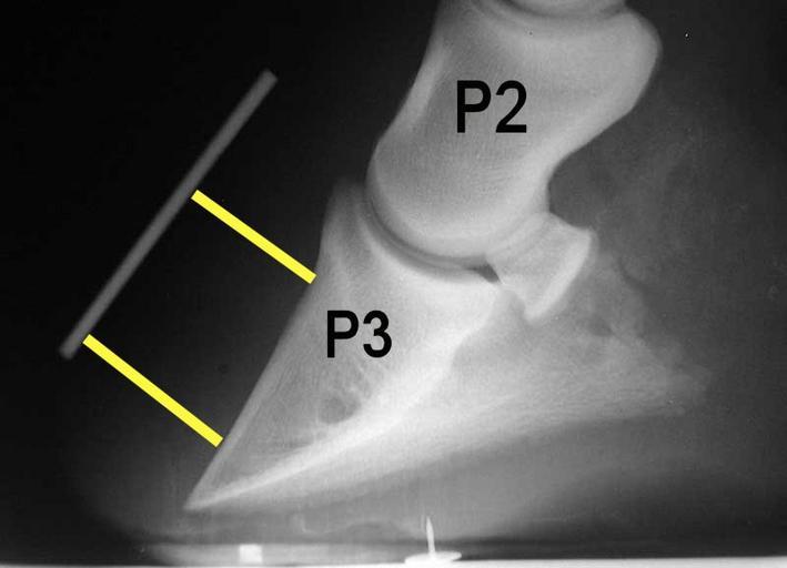
| Keywords: Laminitis radiograph with annotation.jpg en A typical x-ray or radiograph of a laminitic foot in a horse The annotation P2 stands for the middle phalanx pastern bone and P3 denotes the distal phalanx coffin bone The white line marks the boundary of the outer hoof wall and the yellow lines show the distance between the top and bottom part of the coffin bone with the outer hoof wall In this example the distal bottom part of the coffin bone is rotated away greater distance from the hoof wall an indication of laminitis Radiograph created by Malcolm Morley annotated by Froggerlaura Malcolm Morley 1999 Laminitis | ||||