MAKE A MEME
View Large Image

| View Original: | Microvilli.jpg (640x448) | |||
| Download: | Original | Medium | Small | Thumb |
| Courtesy of: | commons.wikimedia.org | More Like This | ||
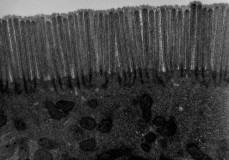
| Keywords: Microvilli.jpg Transmission electron microscope image of a thin section cut through a human jejunum segment of small intestine epithelial cell Image shows apical end of absorptive cell with some of the densely packed microvilli that make up the striated border Each microvillus is approximately 1um long by 0 1um in diameter and contains a core of actin microfilaments http //remf dartmouth edu/images/humanMicrovilliTEM/source/1 html Louisa Howard Katherine Connollly - Dartmouth Electron Microscope Facility http //remf dartmouth edu/imagesindex html http //remf dartmouth edu/imagesindex html Dartmouth Electron Microscope Facility Common Good <span class signature-talk >talk</span> 18 28 13 October 2011 UTC page is was here 18 51 28 December 2002 Magnus Manske 350x247 19 689 bytes <nowiki> Source and public domain notice at http //remf dartmouth edu/imagesindex html </nowiki> Transmission electron microscopic images Histology Animal cells | ||||