MAKE A MEME
View Large Image

| View Original: | Rhodopsin.jpg (622x831) | |||
| Download: | Original | Medium | Small | Thumb |
| Courtesy of: | commons.wikimedia.org | More Like This | ||
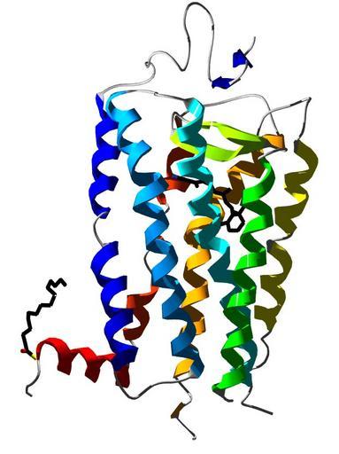
| Keywords: Rhodopsin.jpg Created with Persistence of Vision 3D structure of bovine rhodopsin Derived from the 2 6 Å crystal stucture of rhodopsin 1L9H Structural informations were obtained from pdb org and modelled using spdbv/POV Ray <br/>Blue TMI <br/>Lightblue TMII <br/>Cyan TMIII <br/>Green TMIV <br/>Yellow TMVV <br/>Organge TMVI <br/>Red-orange TMVII <br/>Red Hx8 own 2007-11-11 S Jähnichen Public domain <gallery> File 1L9H Bovine Rhodopsin 2 png File Rhodopsin jpeg </gallery> S Jähnichen Rhodopsins | ||||