Click Here for More Images from iStock- 15% off with coupon 15FREEIMAGES 
Anatomy of the pelvis bones, including the ilium, ischium, sacrum, and pubis, photorealistic 3D illustration. Front view. Perfect for educational or medical purposes.
Mother and daughter putting hands together
Three views of a left hand, the bones, the muscles and tendons, and the flesh epidermal covering
Computational tomography image of the abdominal and pelvic regions.
The annular ligament is a complex structure formed from the capsule, lateral collateral ligamentous complex, and supinator muscle that act in unison to stabilize the proximal radioulnar joint 3d illustration
X-ray image of the hands of a senior woman with Osteoarthritis. Human skeleton.
Carpal bones close-up with body contours 3D rendering illustration isolated on white with copy space. Human skeleton, hand and wrist anatomy, medical diagram, osteology, skeletal system concepts.
MRI scanning of the human body skeletal, muscular and nervous systems with abdomen region view. / You can see the animation movie of this image from my iStock video portfolio. Video number:
Woman hand reaching gesture on white background.
x-ray of the hands, detail of the phalanges and joints,trapeze,scaphoid,Pyramidal,Pisiform,capitate, distal, frontal, proximal, interflaginian, lunate, intercapitate
Fingers developed Raynaud's disease symptoms by cold stimulation.
Dupuytren's contracture after fascioctomy - surgery on right hand little finger of 53 year old man's palm after 40 days. Area was already treated with radio therapy 3 years ago beside needle aponeurotomy 2 years ago . Model released.
The human foot is a highly developed, biomechanically complex structure that serves to bear the weight of the body as well as forces many times the weight of the human body during propulsion.
X-ray of a human female foot, ankle and lower leg
Posterior atlanto axial membrane 3d medical
Hand Gesture
Hand wrinkles
Calcaneus bone of the foot. Heel bone, the largest of the tarsal bones in the foot. Human foot anatomy. 3D illustration. Set of foots in different projections
Mediastinal view of a hamartoma tumor on the left lung. Medical themes
Computed MRI image of Prostate Cancer.
human skeleton anatomy foot bones for medical concept 3D illustration
Inner side of International sign..
The lumbar vertebrae are, in human anatomy, the five vertebrae between the rib cage and the pelvis .
Dupuytren's contracture on right hand little finger of 53 year old man's palm.
Female manicure. Before and after. Girls hands on a white background.
Food Safety with Chicken shaped like a heart on Plastic Cutting Board, protective disposable gloves
Labeled human anatomy Erector Spinae back muscles in isolation on the skeletal system from a posterior view on a white background.  3D rendering.
Female hand holding something. White background.
Cuneiform tarsal bones in red with body 3D rendering illustration isolated on white with copy space. Human skeleton and foot anatomy, medical diagram, osteology, skeletal system concepts.
Radiology of left and right hand
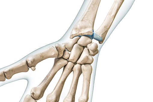
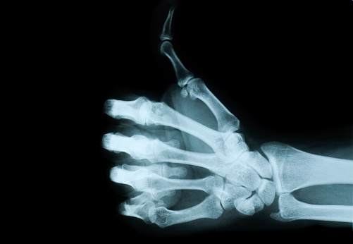
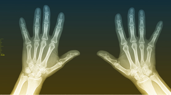
Free Images: "bestof:...Carpals - Trapezoid bone.png Bones of human right hand with thumb on bottom Posterior surface dorsal surface Trapezoid bone labelled File RightHumanPosteriorDis..."
Terms of Use   Search of the Day