Click Here for More Images from iStock- 15% off with coupon 15FREEIMAGES 
A ball and stick model of Human Insulin. Isolated on black.
3D cartoon and Gaussian surface models, chain id color scheme, PDB 2y73, white background
Squalane is a hydrocarbon derived by hydrogenation of squalene. 3d illustration
Covid-19 vaccination record card with fictitious text
Copper(II) sulfate with molecular structure. Chemical ingredient used in medical and public health issues.
Inside protein molecule
3d Rendering Molecular Structure,Ozone,Hydrogen and oxygen
Colonies of Candida Albicans growth on Chocolate agar from sputum culture
Solar radiation analysis of a public building. Multipurpose common public space analysis with parametric louvers to control sun exposure and radiation.
Chemical formula of Adrenaline
3D cartoon and Gaussian surface models, PDB 7bg9, white background.
3d rendering of transparent molecule shape on green color background.
Calcidiol molecule, also called calcifediol, major circulating metabolite of vitamin D3 produced in liver, 3D illustration
nanotechnology and abstract graphene structures
Baryte or barite is a mineral consisting of barium sulfate. The baryte group consists of baryte, celestine, anglesite and anhydrite. 3d illustration
Molecular structure formula. Concept. 3D Render
Water (Molecular formula: H2O) is a clear, odorless, tasteless liquid that is essential for most animal and plant life. Chemical structure model: Ball and Stick. 3D illustration.
Chemical formula of Progesterone
3D cartoon and Gaussian surface models, PDB 7qqf, white background
A model of a molecule of arginine, an amino acid. Amino acids are the building blocks of proteins and have many functions in metabolism. In particular, Arginine has an important function in the immune system, wound healing and cell division.
Glucose-6-phosphate dehydrogenase (G6PD) protein. Enzyme of the pentose phosphate pathway that generates co-enzyme NADPH. 3D illustration.
Crystal Lattice
Closeup chemical ingredient on white laboratory table. Sodium Thiosulfate in chemical watch glass with molecular structure Na2S2O3. Side View
Radiology report. On the right, lateral breast there are two intramammary lymph nodes with fatty hilum measuring 0.5 and 0.6 cm respectively. No suspicious breast mass or axillary lymphadenopathy. Moderately dense breasts with 50 to 75 % glandular tissue.  Bi-RADS 2 : benign findings.
Remdesivir is a broad-spectrum antiviral medication, it is a specific treatment for COVID-19. 3d illustration
Energy exchange of electrons between atoms
3D cartoon and Gaussian surface models, PDB 2vtb, white background.
Surface photography of nature pattern Crystal shot for background.
Molecular model of insulin hormone, 3D illustration. It is produced by pancreas and takes part in glucose and lipid metabolism
Close-up shot of a petri dish for checking an antibiotic in the laboratory.
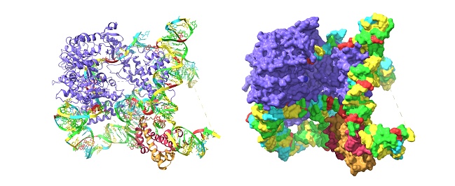
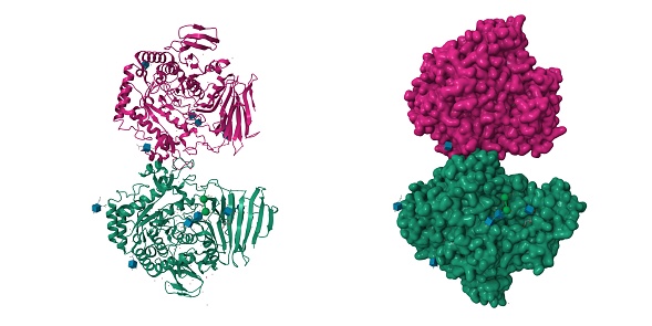
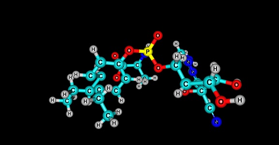
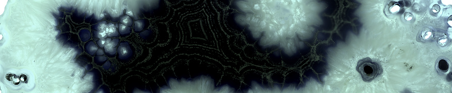
Free Images: "bestof:1pn2.png en Crystal structure analysis of the selenomethionine labelled 2-enoyl-CoA hydratase 2 domain of Candida tropicalis multifunctional enzyme type 2 Pymol"
Glycogen structure.svg
P450cycle.svg
Tetracycline ABCD.svg
Tetracycline.svg
Ergolin num ABCD.svg
Ergolin num.svg
Ergolin.svg
Tetracycline ABCD num.svg
Dynamic array.svg
Work function mismatch gold aluminum.svg
Notable mutations.svg
portugues-sexo_alemão-sex_ingles-sex_africano-seks_russo-секс_albanes-sex_chines-性別_bulgaro-секс_croata-spol_gales-rhyw_coreano-섹스_frances-sexe_japones-セックス_filandes-sukupuolesta_arabe-الجنس_prontos_ai_tens_em_15_linguas_a_p.jpg
1pn2.png
PDB 1pn2 EBI.jpg
ExonucleaseIIIfromEColi.png
1bda.png
EcoRV Crystal Structure.rsh.png
Carbon lattice diamond.png
1NBF-KGramatikoff.jpg
CD86 structure.gif
Crystal structure of SopE.png
Zp3 structure 3d4g.png
RecBCD crystal structure.jpg
NH2-Domäne von Kollagen Typ IX, alpha 2.png
Somatotropin 1HGU.png
Streptokinase 1BML.png
Fructose 1,6-bisphosphatase 1FTA.png
CD4+MHCII 1JL4.png
Asparaginase2 Ecoli 3ECA.png
Apobec.J.Steinfeld.D.png
IL8 Crystal Structure.png
Glycogen structure.png
Helicobacter Pylori Urease.png
EcSHMTPyMol.png
CTLA4 Crystal Structure.jpg
IL19 Crystal Structure.png
IL6 Crystal Structure.png
Neuroligin4.png
GlycogenSyn Wiki.png
PDB 1pn4 EBI.jpg
PDB 1s9c EBI.jpg
1T8U.jpg
3edg.png
ECOR1 Crystal Structure.rsh.png
Ornithine Decarboxylase Publication View.png
The crystal structure of the GPIbα N-terminal domain depicting important ligand binding sites.png
Chain_A,_Crystal_Structure_Of_Mcpip1_Conserved_Domain_With_Zinc-finger_Motif.png
Occludin.png
TlI structure.png
3KSV animation.gif
IL1a Crystal Structure.png
1FMK (c-Src).png
Bcr abl sti.jpg
RXRalpha-alitretinoin.png
Estrogen receptor+lasofoxifene.png
RAP1-rasmol.GIF
Terf2 and tin2.JPG
2B9R.png
Breithauptite structure.png
IL22 Crystal Structure.png
Crystal Structure of Human Diamine Oxidase.png
Crystal_structure_1E3P.jpg
1st2.png
Fumarase 3e04.png
CD32ray.png
Perovskita-wiki.jpg
Iceviistructure-ru.gif
Bcl-xl.jpg
4f0a bio r 500.jpg
Calcineurin 1AUI.png
GIRK 1N9P.png
1I3V (Lama VHH domain unligated).png
1L9H (Bovine Rhodopsin) 2.png
Ribbon diagram depicting the crystal structure of the GPIbβ extracellular domain.png
GNRH 1YY1.png
LDLR 1N7D.png
PDB-2RGT-Complex Lhx3 and Isl1.png
Vts1P SRE Recognition SAM domain.jpg
Subtilisin 1AF4.png
HindIII 2E52.png
PEPCK 1KHF.png
Pyruvate-decarboxylase-1PVD.jpeg
Mutated EGFR kinase domain in complex with gefitinib.png
CBX1+EMSY 2FMM.png
NRas 3CON.png
Pepsin+Pepstatin 1PSO.png
Glutathionperoxidase 2F8A.png
Ovalbumin 1OVA.png
UROD 1URO.png
PSA 2ZCK.png
Urease 2KAU.png
Factor XI 2F83.png
Shiga toxin B 1DM0.png
Shiga toxin 1DM0.png
2FO0 (c-Abl inactive).png
GCL Ecoli 1V4G.png
Crystal Structure of the FokI endonuklease - PDB-2FOK.png
1APS PDB PROTEIN.PNG
Terms of Use   Search of the Day