Click Here for More Images from iStock- 15% off with coupon 15FREEIMAGES 
A ball and stick model of Human Insulin. Isolated on black.
orange: RNA primer; red: RNA template; light blue: proofreading riboexonuclease; mid blue: helicase; dark blue: RNA directed RNA polymerase; violet: non structural proteins (NSPs).
3D cartoon and Gaussian surface models, PDB 6vg8, white background
molecular structure digital concept
Proteasomes are molecular machines for breaking down proteins called proteolysis, only target proteins that have been marked for destruction called ubiquitin to the target protein, 3d rendering
Inside protein molecule
3d Rendering Molecular Structure,Ozone,Hydrogen and oxygen
Multicolored antibodies or immunoglobulin protein structures - 3d illustration
Biological protein and molecule, 3d rendering. 3D illustration.
6G, 6 Generation, Mobile Network Data Technology, Global Communication, Speed
3d illustration of starch (helical amylose) biomolecule, polymer of glucose subunit, main energy storage compound in plants and principal carbohydrate in human diet. Isolated on black.
29 October Republic Day of Türkiye text on a red shiny sphere with a partial Turkish flag on white background. Easy to crop for all your print sizes and social media needs.
PETase is a bacterial enzyme that breaks down PET-plastic to monomeric molecules. The whole bacterial degradation process yields terephtalic acid and ethylene glycol, which are environmentally harmless.
3d render 15 Percent Symbol sitting on Business and Financial and Technical Data Chart (Depth of field)
Water (Molecular formula: H2O) is a clear, odorless, tasteless liquid that is essential for most animal and plant life. Chemical structure model: Ball and Stick. 3D illustration.
Close-up shot of a petri dish for checking an antibiotic in the laboratory.
Calcidiol molecule, also called calcifediol, major circulating metabolite of vitamin D3 produced in liver, 3D illustration
Mobile Network Data Technology, Global Communication, Speed
isolated Chitin is a long-chain polymer of N-acetylglucosamine molecules from polysaccharides family black background 3d rendering
Fictitious molecule model inside red and blue pill isolated on white.
The Raf/MEK/ERK (MAPK) propagates growth signals in the cell. Misregulation can lead to cancer.\nThe red dots on the active EGFR are phosphorylated tyrosines. PDB entries: 1egf, 1nql, 1m17, 2jwa, 3njp, 2gs6, 1gri, 1xd2, 3ksy, 5p21, 6xi7, 6q0j, 2y4i,
Molecular model of Adenosine triphosphate,  or Adenosine-5'-triphosphate (ATP)
3D cartoon and Gaussian surface models, PDB 3hg3, white background
molecular structure digital concept
Biological protein and molecule, 3d rendering. 3D illustration.
6G, 6 Generation, Mobile Network Data Technology, Global Communication, Speed
3d structure of Lactobionic acid, a sugar acid. It is a disaccharide formed from gluconic acid and galactose. It can be formed by oxidation of lactose.
Hong Kong - August 9, 2023 : The Hong Kong Science Park in Pak Shek Kok, New Territories, Hong Kong.
Molecular model of insulin hormone, 3D illustration. It is produced by pancreas and takes part in glucose and lipid metabolism
Palace of the council of Europe in Strasbourg, France. July, 12th, 2020
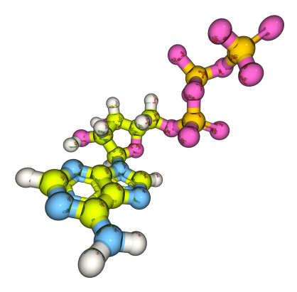
Free Images: "bestof:2C-P molecule 2D.svg 2C-P 2D molecule Own 2007-11-19 Harbin <gallery>File 2 5-Dimethoxy-4-propylphenethylamin svg</gallery> 2C-class phenethylamines"
2C-P molecule 2D.svg
2C-SE molecule 2D.svg
2C-TFM molecule 2D.svg
2C-T molecule 2D.svg
2C B molecule 2D.svg
2C-C molecule 2D.svg
2C-D molecule 2D.svg
2C-H molecule 2D.svg
2C-O molecule 2D.svg
2C-E molecule 2D.svg
2C-G molecule 2D.svg
2C-C-NBOMe.svg
2CBCB-NBOMe.svg
2CBFly-NBOMe.svg
2C-I molecule 2D.svg
2C-TFM 2D.svg
2C-N.svg
2C-O-4.svg
2C-F.svg
2C-T-13.svg
2C-T-17.svg
2C-YN skeletal.svg
2C-TFM-NBOMe.svg
2C-H.svg
2C-E.svg
2C-B.svg
2C-T-7.svg
2C-C.svg
2C-T-2.svg
2C-D.svg
2C-O.svg
2C-P.svg
2C-T.svg
2C-T-4.svg
2C-T-8.svg
2C-T-9.svg
2C-G-5.svg
2C-G.svg
2C-G-N.svg
2C-G-3.svg
2C-G-4.svg
2C-T-21.svg
2C-SE.svg
2C-TFM.svg
2C-T-15.svg
2C-B molecule.svg
2C-E molecule.svg
2C-T-7 molecule.svg
2C-T-21 molecule.svg
Mirtazapine 2D molecule.svg
2C-G-5 2D.svg
2C-B 2D Structure.svg
2C-I.svg
2C-B-BZP.svg
Prenylamine.svg
TFMFly.svg
25I-NBMD.svg
25i-NBF.svg
2C General Structure.svg
2C-I molecule.svg
Cyclohexylacetone 2D.svg
Metaphit.svg
Methamphetamine-2D-skeletal-.svg
Mephentermine.svg
Norpropylhexedrine.svg
Mescaline.svg
Dexfenfluramine.svg
DSP-4.svg
2ct15.svg
2ct17.svg
Amosulalol.svg
Tiflorex.svg
4-D.svg
Mexacarbate-Skeletal-2D.svg
Sulfur-trioxide-2D.svg
Sulfur-monoxide-2D.svg
Thyronamine.svg
Cafedrine 2D SVG.svg
Ganesha.svg
3-Iodothyronamine.svg
3C-BZ-2D-Skeletal.svg
H2O 2D labelled.svg
Indole 2D numbered.svg
Indole 2D aromatic.svg
Benzofuran 2D numbered.svg
Benzofuran 2D aromatic.svg
Chemfm furantetracarboxylic anhydride.svg
Chemfm benzoquinonetetracarboxylic anhydride.svg
Thiomersal-Skeletal-Structure-SVG.svg
Merbromin-Skeletal-Structure-SVG.svg
R-Bromo-DragonFLY.svg
Indole 2D aromatic full.svg
Benzofuran 2D aromatic full.svg
Sulfur-dioxide-resonance-2D.svg
Methane-2D.svg
2C-B-FLY.svg
Pentobarbital 2d.svg
2C-EF.svg
3C-P 2d-skeletal.svg
Alfentanil-2D-skeletal.svg
Terms of Use   Search of the Day