Click Here for More Images from iStock- 15% off with coupon 15FREEIMAGES 
Medical computed tomography or MRI scanner. Back view of blonde nurse sitting at computer, doctor standing, making MRI. Specialists working in clinic. Concept of modern diagnostics.
Female patient getting a CT scan at the hospital - medical exam concepts
Doctor with x-ray of lungs in hands in modern medical radiology hospital. The doctor analyzes the condition of the lungs. The virus attacks the lungs. COVID-19 and medical examinations.
Magnetic resonance images of the brain (MRI brain). Computed resonance imaging of the human brain.
Collection of  MRI heart or Cardiac MRI ( magnetic resonance imaging ) of heart  showing heart beating for detecting heart disease.
Senior radiologist of Oncology institute is examing MRI and CAT scans of human abdomen on his monitors
normal abdomen CT scan
Latin American doctor working at a clinic and looking at a dental x-ray on the computer - healthcare and medicine
Medical computed tomography or MRI scanner. Back view of male doctor sitting at computer, nurse making MRI. Focus on nurse. Specialists working in hospital, studying results, examining.
Closeup of a technician preparing a male patient for an MRI cranial scan.
Radiology
Male doctor examining x-ray.
MRI heart or Cardiac MRI ( magnetic resonance imaging ) of heart compare RVOT and LVOT for diagnosis heart disease.
X-ray of the human mouth showing dental
Radiologist, doctor examining x-ray images of patient for diagnosis. Healthcare, people concept
Cancer treatment in a modern medical private clinic or hospital with a linear accelerator. Professional young doctor \nexamining the results of radiotherapy
Concentrated female doctor examining MRIs in hospital
Lateral view of chest CAT- SCAN that shows popcorn calcification a hamartoma tumor on the left lung. Medical themes
female doctor in white coat x-ray virtual reality glasses technology hospital. High quality photo
Computed tomography of chest organs. CT scan. CT Abdomen Cross section View.
MRI heart or Cardiac MRI ( magnetic resonance imaging ) of heart in Short axis  view showing heart beating of  2 chamber for detecting heart disease.
Side view of senior doctor examining x-rays. Male surgeon is looking at medical report in hospital. He is wearing blue scrubs.
normal head  scan brain window
A young adult female radiographer instructs a young man who is preparing to have a CT scan. The radiograper talks to the man from the CT scan control room.
An EXCELLENT medical scan taken by a radiologist of a horse's right hoof; imagery was required to make proper diagnosis and determine the extent of the mare's injuries.
Doctor looking at his patients X-ray image on digital tablet
Chest CT SCAN of female patient with breast protesis and lung infection
X-ray of female hips, pelvis and spine
Male doctor examining x-ray.
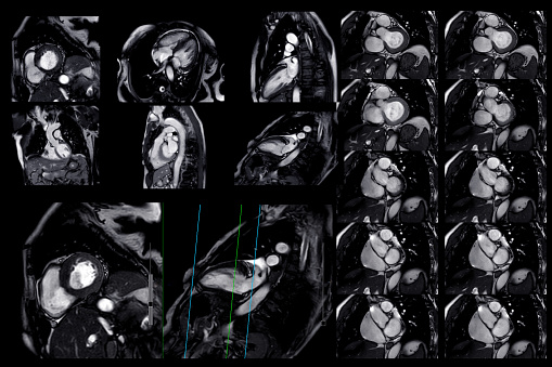
Free Images: "bestof:AFIP-00405558-Glioblastoma-Radiology.jpg en CNS GLIOBLASTOMA MULTIFORME As seen here by magnetic resonance imaging the glioblastoma multiforme usually exhibits"
T2 relaxation.svg
MRI Phase Encoding Principle.svg
MR conditional sign.svg
MR safe sign.svg
AFIP-00405558-Glioblastoma-Radiology.jpg
AFIP00405522M-GLIOBLASTOMA_ARISING_IN_ASTROCYTOMA.jpg
AFIP-00405589-Glioblastoma-Radiology.jpg
AFIP-00405564-Glioblastoma-Micro.jpg
AFIP-00405573-Glioblastoma-Micro.jpg
AFIP-00405590-Glioblastoma-Radiology.jpg
AFIP-00405562-GiantCellGlioblastoma-Gross.jpg
Glioblastoma_multiforme.jpg
FMRI.jpg
Amgydala.jpg
Lascar.jpg
Dgemric cart.jpg
Thalamus2008.jpg
US_Navy_030819-N-9593R-228_Civilian_technician,_Jose_Araujo_watches_as_a_patient_goes_through_a_Magnetic_Resonance_Imaging,_(MRI)_machine.jpg
MRI-DTDA.psd.jpg
MRI-STDA.psd.jpg
T1 Map of cartilage.jpg
Mag suscp phantom.png
Caudate nucleus.jpg
Pallidus.jpg
Precuneus.png
Corps calleux Sagittal.JPG
Magnevist Bottle.JPG
Amygdala Decety.jpg
MRI Location Amygdala up.png
Striatum Structural MRI.png
Substantia Nigra.jpg
Putamen structuralMRI.png
Cuneus.png
MRI Location Hippocampus up..png
Nucleus accumbens sag.jpg
405518-FIBRILLARY_OR_DIFFUSE_ASTROCYTIC_NEOPLASM.jpg
MULTIPLE_SCLEROSIS.JPG
SPINAL_CORD_BLEED.GIF
STENOTIC_CANAL.JPG
Caudate Nucleus Structural MRI.png
Globus Pallidus sturctural MRI.png
Cardiomr dcm.jpg
AnisoColor.tif
Putamen.jpg
HerniatedSpinalDisk-NarrowedNerveRoot.JPG
SPINAL_STENOSIS.JPG
HerniatedSpinalDisk-NarrowedNerveRoot-Contrasty.JPG
1 MRI-Virtual-Window.jpg
MI fMRI.jpg
US_Navy_030821-N-9593R-038_View_of_the_MedModeler.jpg
Varian4T.jpg
Lagehernia.png
US_Navy_030821-N-9593R-044_The_MedModeler_lays_down_a_brown_supportive_base.jpg
US_Navy_030822-N-9593R-008_a_three_dimensional_full-scale_model_of_part_of_a_patient's_face.jpg
NIMA 2A2B3A3B13.png
US_Navy_030820-N-9593R-016_Close-up_view_of_the_MedModeler_at_the_National_Naval_Medical_Center_in_Bethesda,_Maryland.jpg
T1t2PD.jpg
100325 RS88UH 11 qc.gif
MRI Slices - 2007 and 2014 of astrocytoma patient - Steven Keating.jpg
OCD_WalterReed_MRI-Sagital-T2.jpeg
OCD_WalterReed_MRI-Sagital-T1.jpeg
Face_recognition.jpg
Researcher-test.jpg
Cardiac_Mri.jpg
Haxby2001.jpg
Head mri animation.gif
Fingertapping experiment DXIII.jpg
Spinal-disc-protrusion-l5.jpg
Terms of Use   Search of the Day