Click Here for More Images from iStock- 15% off with coupon 15FREEIMAGES 
Green algae cells background
Particles, medical concept
Abstract black and white background picture for computer desktops
Bone marrow plasma cells .Plasma cell neoplasms are diseases in which the body makes too many plasma cells. Plasma cell neoplasms can be benign or malignant. Diagnosed with Immunostain CD138 positive.
Bronchitis and alveolitis in lung of horse infected with equine influenza virus, 200x magnification.
Photomicrograph of two rotifers. Rapidly rotating cilia at top of heads, internal organs visible. Live specimen. Wet mount, 10X objective, transmitted brightfield illumination.
centropyxis aculeata (amoeboid organism) under the microscope - optical microscope x400 magnification
corn stem micrograph with dye
Epidermis. Electron microscope micrograph showing a keratinocyte of spinous layer. The epithelial cell has a polygonal shape, central nucleus with nucleolus, cytoplasm full of keratin filament bundles, and numerous dark desmosomes crossing the intercellular spaces.
plant Pumpkin ovary under light mircoscope with white background
Blue colored stem cells of a lentil plant under the microscope.
Hepatocellular adenoma (HCA; also termed hepatic adenoma) is an uncommon solid, benign liver lesion that develops in an otherwise normal-appearing liver. Typically, HCAs are solitary and are found in young women in association with use of estrogen-containing medications.
Normal red blood cells Under the microscope
Abstract particles
Nodularia sp. toxic algae under microscopic view, nitrogen-fixing cyanobacteria, cyanotoxin, harmful to humans, x40
molecular structure digital concept
Strongyloides stercoralis is a human parasitic roundworm causing the disease strongyloidiasis.
plant rape flower cross section under light mircoscope with white background
Bacteria Enterococcus isolated on white background, 3D illustration. Gram-positive cocci which cause infant endocarditis and other infections
Blastomycosis is a fungal infection caused by inhaling Blastomyces dermatitidis spores. If it involves only the lungs, it is called pulmonary blastomycosis. Only about half of people with the disease have symptoms, which can include fever, cough, night sweats, muscle pains, weight loss, chest pain, and feeling tired.
Freshwater Closterium algae (unicellular charophyte green algae) - optical microscope x200 magnification
Photomicrograph of filamentous blue-green algae, Oscillatoria species. Each band along the stand is one cell. This filament is very short probably having recently parted from a longer one. Filaments are typically many times longer. This algae is mobile, it moves and twists through the water. San Francisco Bay, California, USA. Live specimen. Wet mount, 40X objective, transmitted brightfield illumination. Note - motion blur of live specimen, very shallow depth of field, chromatic aberration and uneven focus are inherent in light microscopy.
Licmophora sp. algae, marine and freshwater diatom under microscopic view. Genus of benthic, photosynthetic and epiphyte diatom
Monkeypox Virus. 3D Render
Backgrounds of Bacteria and Mold under the microscope in Lab.
Different germs in the human intestines called microbiome,Bacteria Lactobacillus in human intestine,Beneficial healthy intestinal bacterium microflora,Gut bacteria
Components of Eukaryotic cell, nucleus and organelles and plasma membrane - 3d illustration
Paraganglioma is a type of neuroendocrine tumor that forms near certain blood vessels and nerves outside of the adrenal glands. The adrenal glands are important for making hormones that control many functions in the body and are located on top of the kidneys.
Cardano Coin (ADA) cryptocurrency 3D Render. Cardano is a decentralised public blockchain cryptocurrency with smart contracts
Close-up shot of a petri dish for checking an antibiotic in the laboratory.
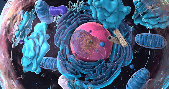
Free Images: "bestof:Animal cell structure numbered version.svg Numbered version of Image Animal cell structure en svg made it myself in adobe ilustrator both jan 2007 original on"
Animal cell structure numbered version.svg
Animal cell structure en.svg
Animal cell structure de.svg
Plant cell structure-en.svg
Diagram cell nucleus no text.svg
Animal cell structure pt.svg
Animal cell structure fr.svg
Animal mitochondrion diagram en.svg
Animal mitochondrion diagram en (edit).svg
Plant cell structure svg labels.svg
Skull template.svg
Average prokaryote cell- en.svg
portugues-sexo_alemão-sex_ingles-sex_africano-seks_russo-секс_albanes-sex_chines-性別_bulgaro-секс_croata-spol_gales-rhyw_coreano-섹스_frances-sexe_japones-セックス_filandes-sukupuolesta_arabe-الجنس_prontos_ai_tens_em_15_linguas_a_p.jpg
Complete neuron cell diagram he.svg
Animal cell structure sv.svg
Animal cell structure id.svg
Animal cell structure no text.svg
Diagram human cell nucleus numbered version.svg
Chloroplast diagram.svg
Animal mitochondrion diagram bn.svg
Prokaryote cell diagram de-2.svg
Plant cell structure svg 2.svg
Prokaryote cell-ro.svg
Zelula prokarioto tipikoa.svg
Average prokaryote cell- nl.svg
Desmosome cell junction en.svg
Przeciętna komórka Prokariotyczna-pl.svg
Average prokaryote cell- lt.svg
Endomembrane system diagram en (edit).svg
Complete neuron cell diagram uk.svg
Cellular tight junction keys.svg
Respiratory system complete ru.svg
Human skull front bones pl.svg
Plant cell structure svg-id.svg
Animal cell structure tr.svg
Animal cell structure pl.svg
Animal cell structure uk.svg
Chloroplast diagram (zh-cn).svg
Complete neuron cell diagram ru.svg
Average prokaryote cell numbered.svg
Animal Cell Unannotated.svg
Average prokaryote cell- fr.svg
Average prokaryote cell cs.svg
Animal mitochondrion diagram de.svg
Sphingolipid de.svg
Flagellum base diagram keys.svg
Average prokaryote cell- unlabled.svg
Mendel seven characters.svg
Prokaryote celle diagram da.svg
Scheme human hand bones-numbers.svg
Beta-thujone.svg
Gas particle movement labeled.svg
2,5-dimethoxybenzaldehyde.svg
Mendel seven characters es.svg
2-hexanol-Line-Structure.svg
Gas particle movement.svg
Mature flower sv blue-labels.svg
Protein primary structure zh (zh-tw).svg
Anisogamy.svg
Insulin glucose metabolism ZP (it).svg
Endomembrane system diagram ka.svg
Os du crane.svg
Complete neuron cell diagram without text.svg
Endomembrane system diagram en.svg
Cochlea.svg
Psilocin structure.svg
Cherokee Syllabary.svg
Adherens Junctions structural proteins-es.svg
Hemi-icosahedron coloured.svg
4-HO-DIPT.SVG
Mature flower diagram cs.svg
Animal mitochondrion diagram cs.svg
Complete neuron cell diagram zh.svg
Complete neuron cell diagram zh-hant.svg
Animal mitochondrion diagram it.svg
Phenibut.svg
Complete neuron cell diagram pt.svg
Average prokaryote cell- ru.svg
Average prokaryote cell- uk.svg
Animal cell structure en gl.svg
Animal cell structure hu.svg
Animal cell structure gl.svg
Animal cell structure ca.svg
Average prokaryote cell- id.svg
IIT-Organisational-structure.svg
Animal cell cycle (editors version).svg
Pentanal.svg
8 Strokes of Han Characters.svg
Plant cell structure fr.svg
Plant cell structure svg pl.svg
Plant cell structure no text-2.svg
Complete neuron cell diagram numbered large numbers.svg
Acrosome reaction diagram es.svg
Point quadtree.svg
Design_Analysis_of_the_B-24J_Liberator.png
Brajlo.svg
Esquelet humà frontal.svg
Terror2.svg
Terms of Use   Search of the Day