Click Here for More Images from iStock- 15% off with coupon 15FREEIMAGES 
3D Rendering of male pelvis, hip, leg bones and ligaments labeled on a white background.  Posterior view.
3D view of a new generation shoe design. The lightness, performance and appearance of the shoes are now indispensable elements of design. The importance of foot health and shoe relationship. / You can see the animation movie of this image from my iStock video portfolio. Video number:
The Plantar aponeurosis is the modification of Deep fascia, which covers the sole 3d illustration
MRI view of ankle.  To see more medical related images:
3D Illustration Concept of Human Skeleton System Bone Joints Anatomy
Foot bones lateral view with body contours 3D rendering illustration isolated on white with copy space. Human skeleton and ankle anatomy, medical diagram, osteology, skeletal system concepts.
Foot x-ray image.
medical accurate illustration of the plantar calcaneonavicular ligament
X-ray of a human female foot, ankle and lower leg
Foot and ankle pain, conceptual 3D illustration. Foot anatomy. Achilles heel pain
MRI scanning of the human body skeletal, muscular and nervous systems with abdomen region view. / You can see the animation movie of this image from my iStock video portfolio. Video number:
Labeled dorsal lateral view of female foot showing bone, muscle and ligaments on white background.
Anatomical Model of a human knee on white background
The lumbar vertebrae are, in human anatomy, the five vertebrae between the rib cage and the pelvis .
Computational tomography image of the abdominal and pelvic regions.
medical accurate illustration of the flexor hallucis brevis
A pair of male feet
For these mild nerve injuries, nonsurgical treatment options include medication, physical therapy or massage therapy. Peripheral nerve surgery can reconstruct or repair damaged nerves. You may need surgery to repair severely compressed nerves, cut nerves or nerves that are not healing on their own.
Computed MRI image of Prostate Cancer.
Mediastinal view of a hamartoma tumor on the left lung. Medical themes
Digital medical illustration: Lateral (side) x-ray view (orthogonal) of human foot and ankle. Featuring:
The flexor retinaculum of the foot extends from the medial malleolus above, to the calcaneus below 3d illustration
Legs and feet of a young woman on a wooden pier in front of the turquoise blue sea in summer
human skeleton anatomy foot bones for medical concept 3D illustration
Man foot on white background
Knee joint exploded view exposing the meniscus connective tissue
Close-up of an acupuncturist's hands giving treatment to a woman of caucasian ethnicity. He is putting the needles in the woman's foot.
The foot (plural feet) is an anatomical structure found in many vertebrates. It is the terminal portion of a limb which bears weight and allows locomotion. In many animals with feet, the foot is a separate organ at the terminal part of the leg made up of one or more segments or bones, generally including claws or nails.
A cgi view of an inflamed joint isolated on blue
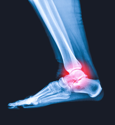
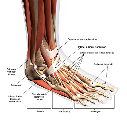
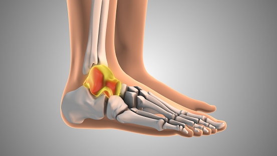
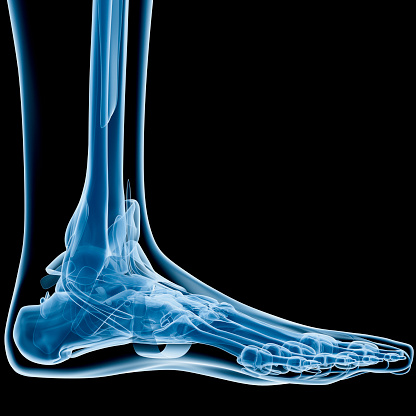
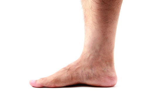
Free Images: "bestof:Ankle.svg Lateral view of the human ankle 1 Fibula 2 Tibia 3 Anterior inferior tibiofibular ligament 4 Anterior tibiofibular ligament 5 Talus 6 Calcaneofibular"
Anterior view of human male, retouched.jpg
Naked human male body front anterior (annotated-en).png
Naked human male body front anterior (annotated-id).png
Naked human male body front anterior.png
Anatomía humana.png
Melissodes_near_bimaculatus,_f,_virginia,_pulaski_county,_face_2016-12-16-13.40.jpg
Melissodes_near_bimaculatus,_f,_virginia,_pulaski_county,_side_2016-12-16-13.52.jpg
Melissodes_near_bimaculatus,_f,_virginia,_pulaski_county,_back_2016-12-16-13.31.jpg
Ankle.svg
Ankle_en.svg
Ankle ar.svg
Ankle_es.svg
Schematic diagram of the human eye multilingual.svg
Circle of Willis en.svg
Anterior view of human female and male, with labels.svg
stubbs_study_human_figure_anterior_view_comparative_anatomical_exposition.jpg
Female reproductive system - anterior view.svg
Anatomia humana.svg
Anatomia czlowieka - nowy.svg
사람의 몸.svg
मानव शरीर.svg
Venous system en.svg
Arterial System en.svg
Circulatory system pl.svg
Circulatory System fr.svg
Venous system fr.svg
Arterial System fr.svg
Circulatory System pt.svg
Anatomía humana - nuevo.svg
Circulatory System en edited.svg
Cubitus varus and cubitus valgus.svg
Anatomie des menschen - neu.svg
Human anatomy in Moksha.svg
Anatomické zobrazení.svg
İnsan anatomisi.svg
Homa korpo.svg
Knee_diagram-de_ACL_PCL.svg
Cos huma davant etiquetes.svg
Circulatory System en.svg
Venous system pl.svg
Arterial system pl.svg
Circulatory System no tags.svg
Circulatory System it.svg
Arterial System es.svg
Venous System es.svg
Circulatory System es.svg
Человеческое тело.svg
Arterial System ru.svg
Circulatory System ru.svg
Vue antérieure de humain de sexe féminin et masculin.svg
Ανθρώπινη ανατομία.svg
Gray512.svg
George_Stubbs_-_A_Comparative_Anatomical_Exposition_of_the_Structure_of_the_Human_Body_with_That_of_a_Tiger_and_a_Co..._-_Google_Art_Project.jpg
George_Stubbs_-_A_Comparative_Anatomical_Exposition_of_the_Structure_of_the_Human_Body_with_That_of_a_Tiger_and_a_Co..._-_Google_Art_Project_(2345851).jpg
George_Stubbs_-_A_Comparative_Anatomical_Exposition_of_the_Structure_of_the_Human_Body_with_that_of_a_Tiger_and_a_Co..._-_Google_Art_Project_(2345889).jpg
George_Stubbs_-_A_Comparative_Anatomical_Exposition_of_the_Structure_of_the_Human_Body_with_that_of_a_Tiger_and_a_Co..._-_Google_Art_Project_(2346871).jpg
George_Stubbs_-_A_Comparative_Anatomical_Exposition_of_the_Structure_of_the_Human_Body_with_that_of_a_Tiger_and_a_Co..._-_Google_Art_Project_(2346882).jpg
George_Stubbs_-_A_Comparative_Anatomical_Exposition_of_the_Structure_of_the_Human_Body_with_that_of_a_Tiger_and_a_Co..._-_Google_Art_Project_(2349560).jpg
George_Stubbs_-_A_Comparative_Anatomical_Exposition_of_the_Structure_of_the_Human_Body_with_that_of_a_Tiger_and_a_Co..._-_Google_Art_Project_(2350908).jpg
George_Stubbs_-_A_Comparative_Anatomical_Exposition_of_the_Structure_of_the_Human_Body_with_That_of_a_Tiger_and_a_Co..._-_Google_Art_Project_(2364017).jpg
George_Stubbs_-_A_Comparative_Anatomical_Exposition_of_the_Structure_of_the_Human_Body_with_That_of_a_Tiger_and_a_Co..._-_Google_Art_Project_(2359178).jpg
George_Stubbs_-_A_Comparative_Anatomical_Exposition_of_the_Structure_of_the_Human_Body_with_that_of_a_Tiger_and_a_Co..._-_Google_Art_Project_(2357268).jpg
George_Stubbs_-_A_Comparative_Anatomical_Exposition_of_the_Structure_of_the_Human_Body_with_That_of_a_Tiger_and_a_Co..._-_Google_Art_Project_(2353350).jpg
George_Stubbs_-_A_Comparative_Anatomical_Exposition_of_the_Structure_of_the_Human_Body_with_That_of_a_Tiger_and_a_Co..._-_Google_Art_Project_(2349702).jpg
George_Stubbs_-_A_Comparative_Anatomical_Exposition_of_the_Structure_of_the_Human_Body_with_that_of_a_Tiger_and_a_Co..._-_Google_Art_Project_(2353489).jpg
George_Stubbs_-_A_Comparative_Anatomical_Exposition_of_the_Structure_of_the_Human_Body_with_That_of_a_Tiger_and_a_Co..._-_Google_Art_Project_(2367079).jpg
Mouvement diaphragme respiration.svg
Esquelet humà frontal.svg
YES_HE_DID_..AND_THIS_IS_WHY.jpg
Lophopsittacus_fossils.jpg
Legamenti crociati.jpg
Ankle.PNG
Gray345.png
Gray326.png
The_Quarterly_journal_of_the_Geological_Society_of_London_(13860274604).jpg
Radiolaria_(Challenger)_Plate_125.jpg
Radiolaria_(Challenger)_Plate_124.jpg
Gray304.png
Gray261.png
Gray146.png
Snowboarders_ankle_-_McCrory.jpg
Gray356.png
Gray737.png
Gray258.png
Greater_sciatic_foramen.png
US_Navy_030710-N-0000W-001_Orthopedics_Department_Head_Lt._Cmdr._Daniel_J._Solomon_performs_exploratory_surgery_during_an_anterior_crucuate_ligament_reconstruction_procedure,_as_Hospital_Corpsman_3rd_Class_Andrew_S._Caldejon_as.jpg
Gray812.png
Tibia (Shinbone) Shaft Fracture.JPG
Gray_737-emphasizing-corpus-callosum.png
Grande_foro_ischiatico.png
Gray202.png
Gray515.png
Gray188.png
Gray140.png
Gray700.png
Gray814.png
Gray196.png
Illu_female_pelvis.jpg
Circle of Willis-hu.png
The_Quarterly_journal_of_the_Geological_Society_of_London_(12711339145).jpg
Terms of Use   Search of the Day