Click Here for More Images from iStock- 15% off with coupon 15FREEIMAGES 
Monkeypox Virus. 3D Render
E coli bacterial culture plate examination in biotechnology/ microbiology laboratory.
Hand with Petri dish or culture media with bacteria on white background with clipping, Test various germs, virus, Coronavirus, COVID-19, Microbial population count, Food science.
Abstract Lactobacillus Bulgaricus Bacteria - 3d rendered microbiology image. Medical research, health-care concept. SEM (TEM)scanning view
Set of Petri dishes with different culture samples on white background, top view
Clean culture of aerobic bacteria on agar plate, biohazard sign in background. Selective focus on petri dish, slightly blue toned, AdobeRGB color profile. Content of my lightbox medical & sciences:
A quadrant streak of the bacterium Staphylococcus aureus cultured or inoculated on Baird-Parker agar with egg tellurite .
Wide composition of various virus cell shapes inside blood plasma
Streptococcus oral Bacteria and Streptococcal infections as gram-positive bacterial outbreak as spherical Streptococcaceae cell division spreading and growing into chains as an infectious disease as streptococcal diseases as strep throat sinus infections or flesh-eating illness.
Bacterial cell. Microscope view. Scientific medical illustration.
Skin microbiome, Healthy, probiotic bacteria on the skin
Disease X Cells. 3D Render
Backgrounds of Characteristics and Different shaped Colony of Bacteria and Mold growing on agar plates from Soil samples for education in Microbiology laboratory.
Close-up shot of a petri dish for checking an antibiotic in the laboratory.
Enterobius vermicularis (EV) eggs. parasite in stool, image under light microscopy 40X objective at medical laboratory.
Double zones can be seen around 3 antibiotic disks. This is a clear sign of more than one bacterium being present. To measure antibiotic susceptibility for each bacterium it will be necessary to isolate E. coli and Klebsiella on separate agar plates.
Bacteria cells isolated on white background with copy space
Intestinal microbiome,Probiotics health benefits,Gut bacteria,lactococcus, propionibacterium, bifidobacterium.
A closeup shot of a laboratory worker examining a green substance on a petri dish while conducting coronavirus research
Bacteria Aggregatibacter, 3D illustration. Aggregatibacter aphrophilus and A. actinomycetemcomitans, Gram-negative bacteria, part of the normal flora of the mouth and throat, also cause endocarditis
Monkeypox Virus. 3D Render
microscopic magnification Legionella pneumophila, Gram-negative bacillus that causes pneumonia. 3d illustration
Scientist examining a microbiological culture
Scientist or doctor hand with a blue glove holding a petri dish with a selective culture of Escherichia coli bacteria
Close-up shot of a petri dish for checking an antibiotic in the laboratory.
Antibiotic resistant bacteria such as MRSA on surface of a Biofilm
In vitro bacterial culture
Multidrug resistant bacteria. Biofilm of bacteria Acinetobacter baumannii, the common causative agent of hospital-acquired infections 3D illustration
Disease X Cells. 3D Render
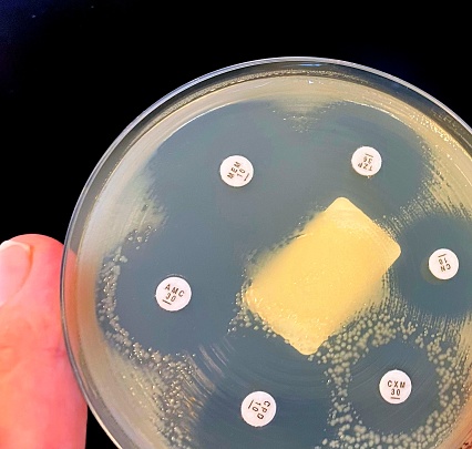
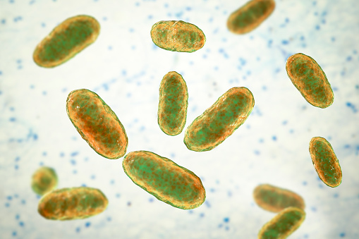
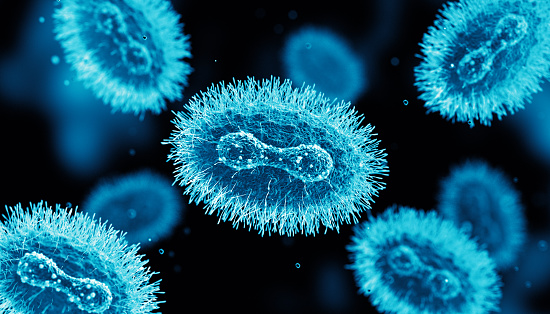
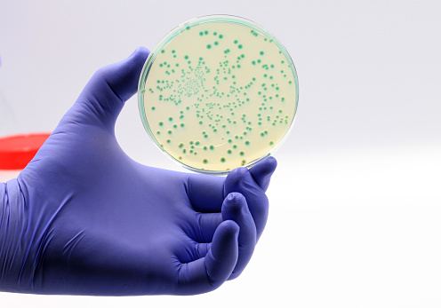
Free Images: "bestof:Bacterial infections and involved species.png en Overview of the main bacterial infections and the most notable species involved See also Wikipedia Bacteria"
Lasioglossum_versatum,_F,_side,_MD,_Cecil_County_2013-07-08-18.19.49_ZS_PMax.jpg
Lasioglossum_versatum,_F,_back,_MD,_Cecil_County_2013-07-08-18.05.57_ZS_PMax.jpg
Lasioglossum_versatum,_F,_face,_MD,_Cecil_County_2013-07-08-18.13.27_ZS_PMax.jpg
Polistes_nest_1,_MD,_side_2015-07-22-17.17.40_ZS_PMax_UDR.jpg
Polistes_nest,_MD,_side_2015-07-22-18.26.08_ZS_PMax_UDR.jpg
Polistes_nest_5,_MD,_side_2015-07-22-18.00.39_ZS_PMax_UDR.jpg
Polistes_nest_6,_MD,_side_2015-07-22-18.12.44_ZS_PMax_UDR.jpg
Polistes_nest_4,_MD,_side_2015-07-22-17.49.59_ZS_PMax_UDR.jpg
Polistes_nest_3,_MD,_side_2015-07-22-17.39.42_ZS_PMax_UDR.jpg
Anthophora_ursina,_M,_Back,_Georgia,_Fannin_County_2014-10-24-15.37.jpg
Anthophora_ursina,_M,_Side,_Georgia,_Fannin_County_2014-10-24-16.21.jpg
Anthophora_ursina,_M,_Face,_Georgia,_Fannin_County_2014-10-24-15.53.jpg
Eucera_dubitata,_m,_ga,_baker,_face2_2014-12-27-20.56.39_ZS_PMax.jpg
Cyclamen_hederifolium,_Aiton_1789_(Ericales:_Primulaceæ:_Myrsinoideæ).jpg
Philæus_chrysops_(Poda_1761)_♂+♀_(Salticidæ:_Plexippinæ:_Hyllini).jpg
Cactus from Macanao Peninsula.jpg
Australian-lizard.JPG
European honey bee extracts nectar.jpg
Megachile_campanulae,_male,_underside_head_2012-07-06-18.44.jpg
Amaryllis_belladona_2.jpg
Megachile_campanulae,_male,_face_2012-07-06-18.26.jpg
Tylopelta_gibbera,f,_full_light_2016-11-12-19.31.jpg
Tylopelta_gibbera,,_f,_sternites_2016-11-12-19.50.jpg
Tylopelta_gibbera,_male_face_2016-11-11-14.jpg
Tylopelta_gibbera,_m,_side_2016-11-11-14.36.jpg
Tylopelta_gibbera,_f,_face_2016-11-12-19.20.jpg
Edwin_Smith_Papyrus_v2.jpg
Groene rups 4-5cm.jpg
Camaecyparis_lawsoniana.jpg
Jacquier_/_Jackfruit.jpg
Caesalpinia_pulcherrima.jpg
Slow Loris Female.jpg
Amaryllis_belladonna.jpg
Gazania_krebsiana.jpg
Groene rups 4-5cm opgerold.jpg
Bacterial infections and involved species.svg
Viral infections and involved species.svg
Viral infections and involved species hy.svg
P450cycle.svg
bacteria-bacterial-species-imitation-108897.jpg
bacteria-bacterial-species-imitation-108898.jpg
bacteria-bacterial-species-imitation-108896.jpg
bacteria-bacterial-species-imitation-106583.jpg
bacteria-bacterial-species-imitation-108895.jpg
bacteria-bacterial-species-imitation-808751.jpg
Meiosis main steps.svg
germ-virus-bug-infection-health-28874.svg
bacteria-pathogen-infection-germs-426998.jpg
bacteria-pathogen-infection-germs-1477522.jpg
bacteria-pathogen-infection-germs-1470777.jpg
bacteria-pathogen-infection-germs-1470778.jpg
bacteria-pathogen-infection-green-426997.jpg
viruses-germs-background-pathogen-1359272.jpg
Bacterial morphology diagram.svg
bacteria-cell-forgery-imitation-1521191.jpg
Flagellum base diagram-fr.svg
Bacterial morphology diagram fr.svg
Neomuratree.svg
Flagellum base diagram en.svg
Bacterial morphology diagram-es.svg
Jellyfish cross section numbered.svg
Bacterial morphology diagram cs (2).svg
Bacterial morphology diagram cs.svg
Mesosome formation.svg
Endocrine caclcium en.svg
Health effects of pollution.svg
Adherens Junctions structural proteins.svg
Bacteria morphologic forms simplified.svg
Flagellum base diagram keys.svg
Adherens Junctions structural proteins-ja.svg
Endocrine caclcium es.svg
Endocrine caclcium it.svg
Main symptoms of Infectious mononucleosis.svg
16S.svg
Prokaryote cell diagram international.svg
Adherens Junctions structural proteins it.svg
ViaCampesinaMembers (October 2008).svg
Effects of sleep deprivation.svg
Locust from the plague in Palestine, 1915.jpg
GHCS JTTtree.svg
Agathis Species Density.svg
Main symptoms of diabetes es.svg
Tetraxylopteris reposana sterile branches.svg
DNA microarray experiment.svg
Health effects of cheese.svg
Symptoms of leukemia.svg
Araucaria Distribtion.svg
Interiör med mor och dotter vid fönster (Ludvig August Smith) - Nationalmuseum - 180227.tif.tiff
Kvinna som flätar sitt hår (Ludvig August Smith) - Nationalmuseum - 180226.tif.tiff
Biological classification S Pengo.svg
Biological classification L Pengo tweaked.svg
En qvindelig Figur, som staar ved en Seng, reder sit Haar (Christoffer Wilhelm Eckersberg) - Nationalmuseum - 180185.tif.tiff
Saurischian & Ornithischian pelvis bone modification w- selected species of Dinosauria.svg
Treaty of Lisbon ratification-accurate.svg
Carapa guianensis distribution.svg
Maximum Likelihood-Tree-Acanthobasidium-Aleurodiscus-Neoaleurodiscus.svg
Wu-UPGMA-Tree-Acanthobasidium-Aleurodiscus-Neoaleurodiscus.svg
Wu-Neoaleurodiscus-Maximum Likelihood-Tree.svg
Phylogenetic_tree-fr.svg
Olvany 4101089538 9c4b2723e3 o.jpg
Terms of Use   Search of the Day