Click Here for More Images from iStock- 15% off with coupon 15FREEIMAGES 
3D cartoon model in two purpendicular projections, PDB 4g0v, white background.
Endosomes - 3d rendered image of Endosomes are membrane-bound organelles. They are parts of endocytic membrane transport pathway.  Endomembrane system in cells. Bilayer Lipid Membrane. Abstract medical illustration. Microscopic view. Microbiologic research concept.
Liposome encapsulation for nanomedicine drug delivery neurodegenerative disease 3d rendering
Digital background depicting innovative technologies in (AI) artificial systems, neural interfaces and internet machine learning technologies
The Raf/MEK/ERK (MAPK) propagates growth signals in the cell. Misregulation can lead to cancer.\nThe red dots on the active EGFR are phosphorylated tyrosines. PDB entries: 1egf, 1nql, 1m17, 2jwa, 3njp, 2gs6, 1gri, 1xd2, 3ksy, 5p21, 6xi7, 6q0j, 2y4i,
Molecular model of fullerene (c60)
Cells in the body examined by endoscope
Close up 3D illustration of microscopic Cholera bacteria infection
3D cartoon and Gaussian surface models, PDB 6kxs, white background
What micro plastic is made of - Chemical ingredients.\nClose up of microplastic on finger concept of water pollution and global warming
antibodies to cell movement of the virus
Endosomes - 3d rendered image of Endosomes are membrane-bound organelles. They are parts of endocytic membrane transport pathway.  Endomembrane system in cells. Bilayer Lipid Membrane. Abstract medical illustration. Microscopic view. Microbiologic research concept.
Main mutation site of the coronavirus variant B.1.531 emerged in South Africa. The spike protein (red) is bound to the angiotensin converting enzyme 2 (blue). The mutation is at the amino acid position 484 (yellow). Source: PDB entry 6cs2.
Covid-19 Coronavirus Variants Omicron Delta Gamma Beta Alpha Pandemic Virus Disease Mutation
DNA close-up, DNA Molecule Model, DNA Strands, DNA animation
3d rendering of Easter eggs background
Cell membrane cholesterol is shown in pink. 3D cartoon and Gaussian surface models, PDB 7rpk, white background.
Evolution and Mystery of Life: Mollusks Living in Water
Poly Protein G with Detection Antibodies to enhance Immunoassays sensitivity on the lipid bilayer bacteria outer layer 3d rendering
A conceptual image of an abstract network of multi-coloured organic strands arranged in a non regular pattern all connected together in a complex manner. The network is made from smaller multi-coloured squares and polyhedra, in vivid colours against a pastel toned background. Shows background and foreground elements to the structure.
Bacterial multidrug efflux pumps (E-CmeB) expel antibiotics  from inside the bacterium (bottom): ciprofloxacin (yellow), chloramphenicol (violet),  ampicillin (red).
DNA A-T Pair
Nucleosome, molecular model. 3D illustration. The nucleosome consists of a DNA double helix wrapped around a core of histone proteins.
background
3D cartoon and Gaussian surface models. PDB 3kqg, chain id color scheme, white background.
Endosomes - 3d rendered image of Endosomes are membrane-bound organelles. They are parts of endocytic membrane transport pathway.  Endomembrane system in cells. Bilayer Lipid Membrane. Abstract medical illustration. Microscopic view. Microbiologic research concept.
Acetylcholine receptors in the lipid layer of cell membrane.
H2 Hydrogen Molecule Fuel Cell Element Vehicle Biodegradable Ethical Renewable Energy
The calcitonin gene related peptide (yellow) binds to its receptor (blue) on neurons and smooth muscle cells of cerebral blood vessels, activating a signal cascade through G-proteins (dark blue) in this cells that  leads to a dilatation of the blood vessels (vasodilatation). Blocking the CGRP receptor diminishes the quantity of migraine attacks.\
plant Leaf of Nerium indicum T.S. under light mircoscope with white background
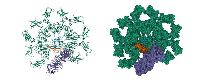
Free Images: "bestof:CD8 receptor.svg en A schematical representation of CD8-receptor found on cytotoxic T cells It consists of an α and a β chain and a carboxy terminus C in the"
CD8 receptor.svg
Antigen_presentation_alt.svg
Convex cone illust.svg
CD8 receptor.PNG
CD8 receptor.png
CD4 receptor.png
PDB 1q69 EBI.jpg
Sickle Cell Anemia.jpg
HLA-A2.png
MHC Class I v2.png
MHC Class II v2.png
LDLR Binding Module5 1ajj.PNG
Total Synthesis of Diltiazem6.png
Determinants of Gastric Acid Secretion.png
Oxygen atmosphere.png
Terms of Use   Search of the Day