Click Here for More Images from iStock- 15% off with coupon 15FREEIMAGES 
From left to right: the NMDA and  AMPA receptors transport calcium cations into neurons after being activated by the neurotransmitter glutamate and  the GABA receptor (right) transport chloride anions after the activation by gamma aminobutyrate. PDB entries: 6wht,  3kg2, 6d6u
Covid-19 Coronavirus Variants Omicron Delta Gamma Beta Alpha Pandemic Virus Disease Mutation
Stomach cancer, symptoms, stages, treatment, 3d illustration
Molecular model of amylose, one of the two sugar components in starch.
Folic acid, folate molecule. It is known as vitamin B9. Molecular model. 3D rendering. Illustration
A molecular model of the flavonoid Quercetin.  An antioxidant and anti-inflamatory, it is used as a nutritional suplement.  It is commonly found in many fruits and vegetables and is a significant chemical component of tea.  Isolated on white.
Tetradecylthioacetic acid molecule. 3D rendering. Atoms are represented as spheres with conventional color coding.
Fictitious molecule model inside red and blue pill isolated on white.
Bacterial multidrug efflux pumps (E-CmeB) expel antibiotics  from inside the bacterium (bottom): ciprofloxacin (yellow), chloramphenicol (violet),  ampicillin (red).
3d Rendering Molecular Structure,Ozone,Hydrogen and oxygen
Deacetoxycephalosporin-C synthase (DAOCS) is a mononuclear ferrous enzyme that transforms penicillins into cephalosporins. 3D cartoon and Gaussian surface models, chain instance color scheme, based on PDB 1uof, white background.
Chemical formula of Progesterone
business strategy concept infographic diagram illustration of balanced scorecard perspectives
Abstract Globe World Maps On White
Chemical formula of Testosterone on a blackboard
3D realistically coloured hexagonal shapes of connected spheres, background with copy space. Futuristic illustration design template for scientific presentation. Molecular business, science, teamwork concept.
After dopamine binding, the dopamine receptor changes his shape and binds the inactive G protein.The G protein expels its GDP molecule replacing it by GTP. and falls into two pieces. The Galpha subunit binds to and activates the enzyme adenylyl cyclase, which produces then cyclic AMP spreading a signal through the cell.
Molecular structure of Sulfur dioxide SO2
Hyaluronan, hyaluronic acid, HA, hyaluronate molecule, short fragment. Molecular model. 3D rendering. Illustration
Molecular model of lactose, a disaccharide sugar in milk.
Nucleosome structure. Structure of an oligonucleosome, showing the packaging of DNA in chromosomes. 3D illustration.
A molecular model of Bisphenol A or BPA.  It is a common building block in the production of many epoxy resins and plastics including polycarbonate plastic. As such it is present in a huge range of food packaging and household products. There are now concerns about its health effects, particularly its effect on children, infants and fetuses.   Isolated on white.
Breast Cross Section of Fat .
Open capsule or pill with molecular structure. Nanotechnology concept
The epidermal growth factor (EGF, red) activates the EGF receptor by binding to it. The activated receptor promotes migration, adhesion and proliferation  of cells. Source: PDB entries, 1 EGF,  1nql, 1ivo, 2jwa, 1m17 and 2gs6.
Chemical formula of Testosterone
B-chain pentamer and A-chain subunit (green), 3D cartoon and Gaussian surface model, white background
Molecular Model of Vitamin E.
Cediranib cancer drug molecule. 3D rendering. Atoms are represented as spheres with conventional color coding.
Crystal Lattice
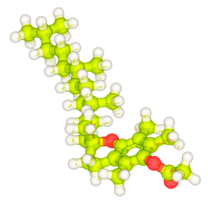
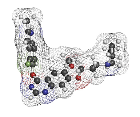
Free Images: "bestof:Carebastine skeletal.svg carebastine the active metabolite of ebastine The difference to ebastine is highlighted own based on File Ebastine svg Anypodetos"
Carebastine skeletal.svg
Sacubitrilat skeletal.svg
Ebastine.svg
Prasugrel active metabolite.svg
Sergliflozin skeletal.svg
Desfesoterodine skeletal.svg
FK1012 skeletal.svg
WR-1065 skeletal.svg
MRE 269 skeletal.svg
Calcium alginate skeletal.svg
Truxinic acid skeletal.svg
Terutroban acid skeletal.svg
Eritoran free acid skeletal.svg
Prasugrel activation.svg
Antibody with CDRs.svg
Ergoloid skeletal.svg
Clopidogrel activation.svg
Emtansine mab structure coloured.svg
Thiouric acid skeletal.svg
Eslicarbazepine structure.svg
Lornoxicam skeletal.svg
SN-38.svg
Polidocanol skeletal.svg
Mertansine skeletal.svg
Capromorelin skeletal.svg
Tabimorelin skeletal.svg
Gitoformate skeletal.svg
Fluclorolone skeletal.svg
Prerapamycin skeletal.svg
Lomitapide skeletal.svg
Eritoran skeletal.svg
R-Isoglutamine skeletal.svg
S-Isoglutamine skeletal.svg
Ibutamoren skeletal.svg
Alpha-cedrene skeletal.svg
Beta-cedrene skeletal.svg
Terutroban structure.svg
Dextronorgestrel.svg
DOTATATE.svg
Dihydrostreptazolin.svg
CI-930 skeletal.svg
Sinay modification.svg
Tetracube branch.svg
Tedizolid phosphate.svg
Dextrin skeletal.svg
Enalkiren skeletal.svg
Enalkiren skeletal 2.svg
Octreotate.svg
SM-130,686 skeletal.svg
MK-5932 skeletal.svg
BIBF 1202.svg
Trifluoromethyl-pyrimidinedione.svg
Ferrier carbocyclization mech.svg
Tetromino I.svg
Tetromino O.svg
Tetromino T.svg
Tetromino J.svg
Tetromino L.svg
Tetromino S.svg
Tetromino Z.svg
Tetracube l-screw.svg
Tetracube r-screw.svg
Catumaxomab mechanism.svg
Catumaxomab structure.svg
Brivudine triphosphate.svg
Ferrier rearrangement.svg
Ferrier carbocyclization.svg
Mertansine mab structure.svg
Sinay transition state.svg
Levofolinic acid.svg
Kavain.svg
Ergotoxine skeletal.svg
Proton pump inhibitors mechanism.svg
CI-930 HPI pattern.svg
Mertansine mab structure coloured.svg
Ferrier carbocyclization 2.svg
Polycarbophil calcium skeletal coloured.svg
Patiromer skeletal.svg
Sumatriptan binding site.svg
Cisplatin synthesis.svg
Colestipol skeletal.svg
Colestipol skeletal 2.svg
Truxillic and truxinic acid stereo.svg
Acemetacin metabolism.svg
Development of nervous system.svg
Bi- and ter-metallocenes containing rhodocenyl groups.svg
Synthesis of cation of ((C5tBu3H2)Rh(C5H5))BF4.svg
River Test map.svg
California county map (Inland Empire highlighted).svg
Bi-specific T-cell engager.svg
Suffolk County, NY, towns and villages Saltaire highlighted.svg
Suffolk County, NY, towns and villages Head of the Harbor highlighted.svg
Saxony C.svg
California county map (Inland Empire highlighted) Gold color.svg
Saxony ERZ.svg
Saxony Direktionsbezirk Dresden.svg
Saxony Direktionsbezirk Leipzig.svg
Saxony Direktionsbezirk Chemnitz.svg
Saxony Z.svg
Saxony V.svg
Terms of Use   Search of the Day