Click Here for More Images from iStock- 15% off with coupon 15FREEIMAGES 
Lipid bilayer cell membrane with membrane and intracellular receptors
Particles, medical concept
3D illustration of sanitary piping
corn stem micrograph with dye
Light bar with LED bulbs  Lighting equipment on white background
Photomicrograph of potato starch in cells of potato tuber, Solanum tuberosum. Starch is normally white but is stained blue with iodine to show better. Live specimen. Wet mount, 10X objective, transmitted brightfield illumination. Note - very shallow depth of field, chromatic aberration and uneven focus are inherent in light microscopy.
The glucagon like peptide-1 receptor (GLPr) is a G proteincoupled receptor (GPCR) that mediates the action of GLP-1, a peptide hormone. The activated receptor  has a strong effect  on the management of type 2 diabetes mellitus and obesity, including glucose homeostasis and regulation of gastric motility and food intake.
Blastomycosis is a fungal infection caused by inhaling Blastomyces dermatitidis spores. If it involves only the lungs, it is called pulmonary blastomycosis. Only about half of people with the disease have symptoms, which can include fever, cough, night sweats, muscle pains, weight loss, chest pain, and feeling tired.
Isolated lipid bilayer coating mesoporous silica nanoparticle 3d rendering in the background
Stem of Nerium indicum T.S. under light mircoscope with white background
Cell Membrane and Molecules, 3d rendering. Computer digital drawing.
Quantum Gravity 3d rendered image of a comprehensive exploration of Quantum Gravity theories. String theory, loop quantum gravity (LOG), causal dynamical triangulation, twistor theory, gravitational waves, spacetime, gravity waves, quantum mechanics, quantum entanglement concepts.
Dicot plants stem show plant vascular tissue under light microscope view for botany education.
Texture Pattern Design on Banknote
Plasma Membrane Of  Cell With other molecules, 3d render
Diffuse large B-cell lymphoma, or DLBCL, is a cancer that starts in white blood cells called lymphocytes.
Leiden leaf c.s. under microscope
plant rape flower cross section under light mircoscope with white background
The study of plant tissues under the microscope in the laboratory.
Oil Tank Wireframe
3d absorbent layer and ventilate shows with synthetic fiber hair, water droplets for diapers, sanitary napkin isolated. baby diaper adult concept, 3d render illustration
Christmas background composition with Christmas baubles on white snow.
Gram-negative, rod-shaped bacteria are a type of bacteria characterized by their appearance under a microscope and their cell wall structure. They have a thin peptidoglycan layer sandwiched between two lipid membranes, which makes them more resistant to certain antibiotics compared to Gram-positive bacteria. Common examples include Escherichia coli (E. coli), Salmonella, and Pseudomonas aeruginosa. These bacteria can cause a wide range of infections in humans, ranging from mild gastrointestinal illnesses to severe systemic infections, and they play significant roles in both environmental and clinical settings.
Captured by a scientific microscope and Canon 5D Mark IV
Glycolic acid is a type of alpha hydroxy acid (AHA). Alpha hydroxy acids are natural acids found in foods. Glycolic acid comes from sugarcane. Alpha hydroxy acids like glycolic acid work by removing the top layers of dead skin cells. Glycolic acid also seems to help reverse sun damage to the skin.
anaplastic thyroid carcinoma with heterologous differentiation (leiomyosarcoma)
Internal structure of an animal cell, 3d rendering. Section view. Computer digital drawing.
Tongue L.S. under light microscope with white background
Mitochondria - microbiology, cellular organelle 3d illustration
Intricate bubble texture macro in bright colors for abstract background. Liquid bubbles lit from below with colors injected into the liquid as a design or art abstract.
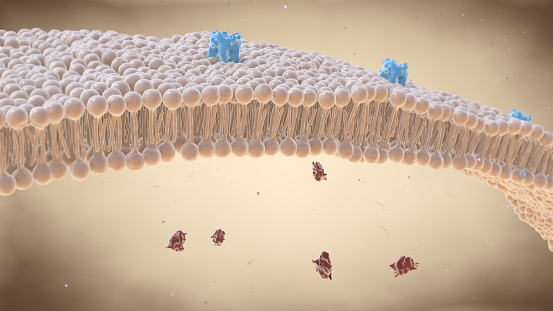
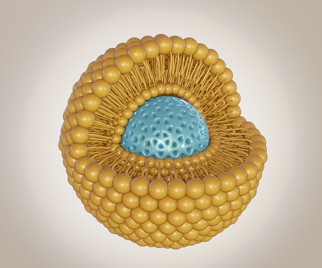
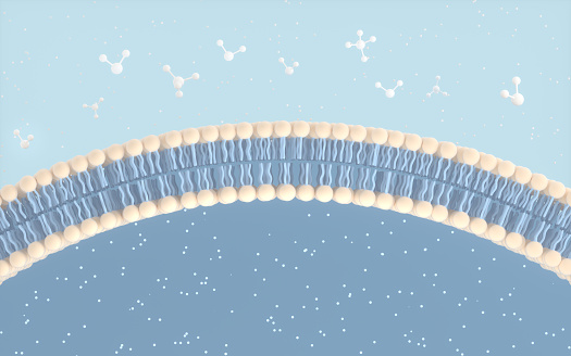
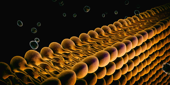
Free Images: "bestof:Cell membrane detailed diagram ua.svg uk Схема будови плазматичної мемб� ани тва� инної клітини 2011-06-23"
Cell membrane detailed diagram ua.svg
Complete neuron cell diagram uk.svg
Average prokaryote cell- uk.svg
Cell membrane detailed diagram es.svg
Cell membrane detailed diagram fr.svg
Cell membrane detailed diagram pt.svg
Cell membrane detailed diagram zh.svg
Cell membrane detailed diagram id.svg
Cell membrane detailed diagram et.svg
Cell membrane detailed diagram de.svg
Cell membrane detailed diagram pl.svg
Cell membrane detailed diagram lv.svg
Cell membrane detailed diagram nl.svg
Cell membrane detailed diagram blank.svg
Cell membrane detailed diagram en.svg
Cell membrane detailed diagram ca.svg
Diagram human cell nucleus uk.svg
Animal cell structure uk.svg
Metabolism diagram.svg
Martello tower diagram UK.svg
Animal mitochondrion diagram uk.svg
Complete diagram of a human spermatozoa uk.svg
Turgor pressure on plant cells diagram-uk.svg
Endomembrane system diagram en (edit).svg
Adherens Junctions structural proteins.svg
Animal mitochondrion diagram bn.svg
Animal mitochondrion diagram en (edit).svg
Animal mitochondrion diagram en.svg
Cell membrane detailed diagram-el.png
Cell_membrane_detailed_diagram.png
Kanalprotein 01.png
GABAB-receptor1.jpg
Plant Microbial Fuel Cell.png
Terms of Use   Search of the Day