Click Here for More Images from iStock- 15% off with coupon 15FREEIMAGES 
Host cells with spores (mold) are inside wood under the microscope for education.
Cirrhosis is a complication of liver disease which involves loss of liver cells and irreversible scarring of the liver. Typically, the disease comes on slowly over months or years. Early on, there are often no symptoms.  As the disease worsens, a person may become tired, weak, itchy, have swelling in the lower legs, develop yellow skin, bruise easily, have fluid build up in the abdomen, or develop spider-like blood vessels on the skin. Cirrhosis is most commonly caused by alcohol, hepatitis B, hepatitis C, and non-alcoholic fatty liver disease. Non-alcoholic fatty liver disease is due to a number of reasons, including being overweight, diabetes, high blood fats, and high blood pressure.  Cirrhosis is characterized by the replacement of normal liver tissue by scar tissue. These changes lead to loss of liver function. Diagnosis is based on blood testing, medical imaging, and liver biopsy.  Image illustrates hepatic steatosis and chronic hepatitis.  Trichrome stain
Microscopic photo of a thin section of calcareous tufa of Holocene age.
Remains of the Roman amphitheatre at the village of Volterra in the Tuscany region of Italy.
Epidermis. Electron microscope micrograph showing a keratinocyte of spinous layer. The epithelial cell has a polygonal shape, central nucleus with nucleolus, cytoplasm full of keratin filament bundles, and numerous dark desmosomes crossing the intercellular spaces.
Photomicrograph of two rotifers. Rapidly rotating cilia at top of heads, internal organs visible. Live specimen. Wet mount, 10X objective, transmitted brightfield illumination.
Natural pools Charcones with green algae in Lanzarote, Canary Islands, Spain
Thracian sanctuary, located near Tatul village, Kardjali region, Bulgaria. Carved grave cut in the rock, II millennium BC.
El Torcal de Antequera es uno de los paisajes Karsticos más emblemáticos de España. Se encuentra en la provincia de málaga, en Andalucía
anaplastic thyroid carcinoma with heterologous differentiation (leiomyosarcoma)
Penicillium branches, ascomycetous fungi under the microscope. To humans ascomycetes are a source of medicinal compounds, like antibiotics, for fermenting bread, alcoholic beverages and cheese. Photo.
Deep wagon wheel ruts in soft rock along the Mormon/Oregon Trail, Wyoming.
Cross-section Dicot, Monocot and Root of Plant Stem under the microscope for classroom education.
Old orange and black scuffed damaged marbled paper, Original over 150+ years old
Breast tissue adenocarcinoma immunohistochemistry. Estrogen receptors.
It is an important historical and archaeological site in Antalya province, Turkey. It exhibits ancient artifacts belonging to the Lycian civilization.
Diatoms are photosynthesising algae, they have a siliceous skeleton (frustule) and are found in almost every aquatic environment including fresh and marine waters, soils, in fact almost anywhere moist.
Carcinoid tumors are a type of slow-growing cancer that can arise in several places throughout your body. Carcinoid tumors, which are one subset of tumors called neuroendocrine tumors, usually begin in the digestive tract (stomach, appendix, small intestine, colon, rectum) or in the lungs. Site: Ovary
Orange and gray lichens on coastal limestone stones and rocks in Crimea, Tarkhankut
Perperikon, Kardzhali region, Bulgaria - August 8, 2020:  Tourists and working people at the Archaeological complex Perperikon. Kardzhali region, Bulgaria, Europe.
Bath in Perge Ancient City in Antalya City, Turkiye
Photomicrograph of midge fly, Chironomidae family, eggs hatching. Eggs are encased in a gelatinous material that attracts debris. Live specimen. Larva about 0.5 mm long. Wet mount, 10X objective, transmitted brightfield illumination.
Hattusa Ancient City old city ruins. Hittite civilization capital Hattusa ruins and architecture. Corum, Turkey
Lichen and moss on rock in New England stone wall, with autumn leaves at edge of frame
Cotinus coggygria bright blossom bush concept photo. Royal purple smoke bush, smokebush, smoke tree and purple smoke tree. High quality picture for wallpaper
Myxoid liposarcoma.  Myxoid/round cell liposarcoma, or MRCLS, is one of several types of liposarcoma. Liposarcoma is a rare cancer that grows in the cells that store fat in the body. MRCLS usually grows in the arms and legs. These tumors grow slowly, and they can spread to other parts of the body
Yellow slime mold, also called dog vomit slime mold, Physarum polycephalum, growing over a fallen log in the Belding Wildlife Management Area of Vernon, Connecticut.
Close-up textured bakcground shot of Weathered damaged pink wall with moss growing
Toxoplasma gondii oocyst under the microscope, isolated
Lembotropis nigricans, also called Cyticus nigricans and Black bloom, is a species of flowering plant in the subfamily of Faboideae of the family Fabaceae. Masses of yellow flowers appear in long racemes in late spring and summer.
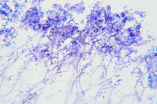
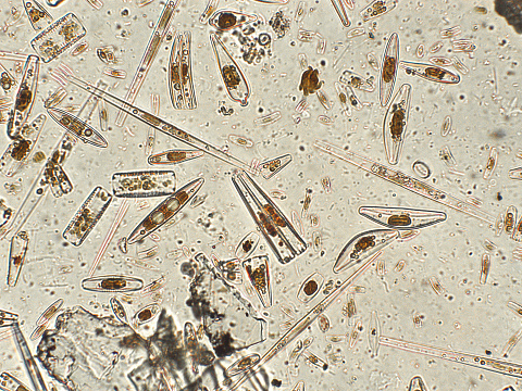
Free Images: "bestof:Conidia and conidiophores of the fungus Acremonium falciforme PHIL 4168 lores.jpg This photomicrograph shows conidia and conidiophores of the fungus Acremonium"
Conidia_and_conidiophores_of_the_fungus_Acremonium_falciforme_PHIL_4168_lores.jpg
Conidiophores_and_conidia_of_the_fungus_Sporothrix_schenckii_PHIL_4208_lores.jpg
Chain_of_conidia_of_a_Alternaria_sp._fungus_PHIL_3963_lores.jpg
Chain_of_conidia_of_a_Alternaria_sp._fungus_PHIL_3962_lores.jpg
Acremonium_falciforme_PHIL_4167_lores.jpg
Conidia-laden_conidiophores_of_Phialophora_verrucosa_PHIL_4238_lores.jpg
Conidiophore_of_the_fungus_Bipolaris_hawaiiensis_PHIL_4338_lores.jpg
Microconidia_of_the_fungus_Trichophyton_mariatii_PHIL_3050_lores.jpg
Macroconidia_of_the_dermatophytic_fungus_Epidermophyton_floccosum_PHIL_4207_lores.jpg
Madurella_grisea_PHIL_4158_lores.jpg
Conidiophores_of_corn_gray_leaf_spot_fungus.jpg
Conidia_phialoconidia_of_Aspergillus_fumigatus_PHIL_300_lores.jpg
Geomyces-007.jpg
Terminal_chlamydospores_of_the_fungus_Microsporum_audouinii_PHIL_3054_lores.jpg
The_fine_branching_tubes_of_the_fungus_Exserohilum_rostratum_PHIL_3784_lores.jpg
Glarea lozoyensis.png
Trichophyton_mentagrophytes_PHIL_2929_lores.jpg
Trichophyton_mariatii_PHIL_3053_lores.jpg
Trichophyton_terrestre_PHIL_4300_lores.jpg
Tuberculate_macroconida_of_the_Jamaican_isolate_of_Histoplasma_capsulatum_PHIL_4023_lores.jpg
Plate_culture_of_Exserohilum_rostratum_PHIL_3786_lores.jpg
4809_lores.jpg
Image_from_page_167_of_"Bacteriological_methods_in_food_and_drug_laboratories,_with_an_introduction_to_micro-analytical_methods"_(1915).jpg
Image_from_page_244_of_"Bulletin_de_la_Société_royale_de_botanique_de_Belgique"_(1911).jpg
Terms of Use   Search of the Day