Click Here for More Images from iStock- 15% off with coupon 15FREEIMAGES 
Mitochondria, a membrane-enclosed cellular organelles producing energy, 3D illustration
What micro plastic is made of - Chemical ingredients.\nClose up of microplastic on finger concept of water pollution and global warming
Host cells with spores (mold) are inside wood under the microscope for education.
3d Mitochondrion isolated on black
Human cell anatomy. 3d illustration
Photomicrograph of filamentous blue-green algae, Oscillatoria species. Each band along the stand is one cell. This filament is very short probably having recently parted from a longer one. Filaments are typically many times longer. This algae is mobile, it moves and twists through the water. San Francisco Bay, California, USA. Live specimen. Wet mount, 40X objective, transmitted brightfield illumination. Note - motion blur of live specimen, very shallow depth of field, chromatic aberration and uneven focus are inherent in light microscopy.
plant Tilia Stem C.S. under light mircoscope with white background
Mitochondria is a double membrane-bound organelle found in all eukaryotic organisms. are related to the cellular respiration process. 3D illustration
Epidermis. Electron microscope micrograph showing a keratinocyte of spinous layer. The epithelial cell has a polygonal shape, central nucleus with nucleolus, cytoplasm full of keratin filament bundles, and numerous dark desmosomes crossing the intercellular spaces.
Natural agate surfaces, suitable for backgrounds and wallpapers.
Cross-section view of Mitochondria, 3d rendering. Computer digital drawing.
Hepatocellular adenoma (HCA; also termed hepatic adenoma) is an uncommon solid, benign liver lesion that develops in an otherwise normal-appearing liver. Typically, HCAs are solitary and are found in young women in association with use of estrogen-containing medications.
The most prominent roles of mitochondria are to produce the energy currency of the cell, ATP (i.e., phosphorylation of ADP), through respiration, and to regulate cellular metabolism.
Golden Turkish Lira Symbol
Leiden leaf c.s. under microscope
plant rape flower cross section under light mircoscope with white background
3d rendering of biological animal cell with organelles cross section isolated on white. Animal cell with placed text annotations to all organelles
Curly marble stone with fashion veins.
Aerial view of loyster farm by Ardara, County Donegal - Ireland.
Photomicrograph of the ciliate Blepharisma americanum in various stages of development. Typical faint pink color. Each individual is only one cell. Live specimen. Wet mount, 10X objective, transmitted brightfield illumination.
Illustration of animal cell anatomy
Microscopic image of dog stomach cardiac region
The intermembrane transporter of the Gram-negative bacterium Acinetobacter baylyi transports a lipopolysaccharide (orange) through the inner membrane consuming ATP (red)
Christmas background composition with Christmas baubles on white snow.
Breast Cross Section of Fat .
Fish gill C.S. under light microscope with white background
Internal structure of an animal cell, 3d rendering. Section view. Computer digital drawing.
Fatty liver disease (steatosis) is a common condition caused by having too much fat build up in your liver. A healthy liver contains a small amount of fat. It becomes a problem when fat reaches 5% to 10% of your liver's weight.
Cells consist of a protoplasm enclosed within a membrane, which contains many biomolecules such as proteins and nucleic acids.
Texture Pattern Design on Banknote
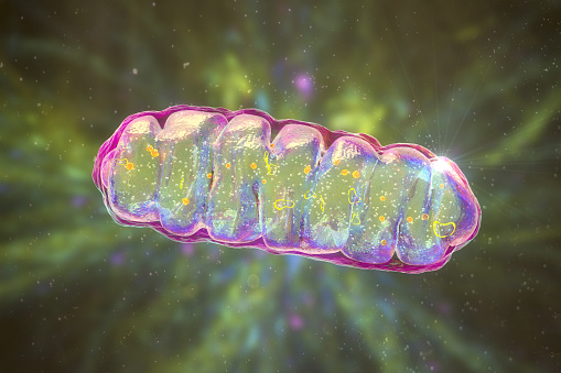
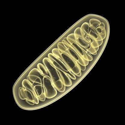
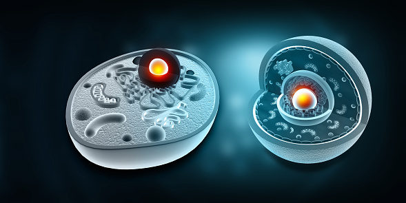
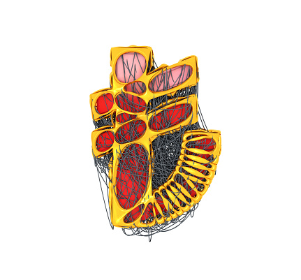
Free Images: "bestof:Diagram of a human mitochondrion blanc.png Diagram_of_a_human_mitochondrion_en svg 2007-12-25 by User Vicki Doronina based on image by Mariana Ruiz"
Diagram human cell nucleus sl.svg
Animal mitochondrion diagram id.svg
Complete diagram of a human spermatozoa pl.svg
Complete diagram of a human spermatozoa pt.svg
Circulatory System en edited.svg
Animal mitochondrion diagram zh hk.svg
Animal mitochondrion diagram zh tw.svg
Animal mitochondrion diagram ml.svg
Animal mitochondrion diagram zh ml.svg
Animal mitochondrion diagram ru.svg
Circulatory System ru.svg
Animal mitochondrion diagram fi.svg
Animal mitochondrion diagram pt.svg
Animal mitochondrion diagram pl.svg
Animal mitochondrion diagram hu.svg
Simplified spermatozoon diagram cs.svg
Diagram human cell nucleus tr.svg
Diagram human cell nucleus (zh-cn).svg
Endomembrane system diagram cs.svg
Diagram human cell nucleus cs.svg
Diagram human cell nucleus pt.svg
Diagram human cell nucleus hu.svg
Diagram human cell nucleus oc.svg
Diagram human cell nucleus id.svg
Human skull front bones pl.svg
Cell junction simplified it.svg
Circulatory System it.svg
Digestive system diagram no labels arrows.svg
Human skeleton front sl.svg
Human skeleton back sl.svg
Cell junction simplified pt.svg
Lebenszyklus der Angiospermen.svg
Human skeleton front ru.svg
Human skeleton back ru.svg
Complete diagram of a human spermatozoa cs.svg
Angiosperm life cycle diagram-no.svg
Human skeleton front af.svg
Endomembrane system diagram ka.svg
Axial skeleton diagram blank.svg
Human skeleton back vi.svg
Esquelet humà frontal.svg
Appendicular skeleton diagram blank.svg
Culex pipiens diagram uk.svg
Culex pipiens diagram es.svg
Pectoral girdle front diagram de.svg
Culex pipiens diagram fr.svg
Pectoral girdle front diagram nan.svg
Arrangement of cocci bacteria cs2.svg
Animal cell structure no text.svg
Animal cell structure id.svg
Animal mitochondrion diagram it.svg
Human skeleton front - no labels.svg
Morphologie bei Bakterien.svg
Prokaryote cell diagram pt.svg
Prokaryote cell diagram es.svg
Human skeleton back no-text no-color.svg
Prokaryote cell diagram no.svg
Human skull side suturas right pl.svg
Bacterial morphology diagram cs.svg
Complete diagram of a human spermatozoa en.svg
Digestive system diagram hu.svg
Digestive system diagram mk.svg
Digestive system diagram sr.svg
Human skull side suturas pl.svg
Human skull side details pl.svg
Gap cell junction-cs.svg
Human leg bones labeled-pl.svg
Mature flower diagram-de.svg
Mendel seven characters cs.svg
Human skull side details right pl.svg
Digestive system diagram da.svg
Fordøjelsen (Dansk).svg
Human skeleton front nan.svg
Human skeleton back nan.svg
Cell junction simplified zh traditional.svg
Mature flower diagram-ca.svg
Filtration diagram (zh-cn).svg
Central nervous system.svg
Prokaryote cell diagram cs.svg
Cell junction simplified cs.svg
Gap cell junction-de.svg
Gap cell junction it.svg
Filtration diagram danish.svg
Connexon and connexin structure cs.svg
Human skull side complete pl.svg
Human leg bones labeled.svg
P Anatomy.svg
Fleur mature français.svg
Filtration diagram (zh-tw).svg
Human skeleton back en.svg
Circulatory System fr.svg
Venous system fr.svg
Plant cell structure svg zh-hant.svg
Human skeleton front en.svg
Scheme human hand bones-ru.svg
Receptive field.svg
Skull template.svg
Scheme human hand bones-numbers.svg
Os du crane.svg
Angiosperm life cycle diagram-en.svg
Terms of Use   Search of the Day