Click Here for More Images from iStock- 15% off with coupon 15FREEIMAGES 
Micrograph of amoeba.  Live specimen. Wet mount, 40X objective, transmitted brightfield illumination. Note - motion blur of live animal, very shallow depth of field, chromatic aberration and uneven focus are inherent in light microscopy.
prostate cancer, prostatic hyperplasia, Structure of male prostate, cross section, 3d illustration
centropyxis aculeata (amoeboid organism) under the microscope - optical microscope x400 magnification
Blastomycosis is a fungal infection caused by inhaling Blastomyces dermatitidis spores. If it involves only the lungs, it is called pulmonary blastomycosis. Only about half of people with the disease have symptoms, which can include fever, cough, night sweats, muscle pains, weight loss, chest pain, and feeling tired.
Backgrounds of Bacteria and Mold under the microscope in Lab.
Squash preparation of stained onion root tip cells showing various stages of mitosis. Differential interference contrast optics on a Leica microscope, Image taken with the X40 objective lens.  Depth of focus is extremely shallow (just a few microns).  Differential interference contrast results in an optical slice horizontally through the specimen providing maximum possible resolution at this magnification and good contrast. Chromatic abberation is inevitable in light microscopy, but has been kept to a minimum in this image.
Liver Fluke Section under light microscope with white background
Implanted human egg of about 10 days. The syncytiotrophoblast form the trophoblastic lacunae. In the center, is the cytotrophoblast. Embryo cells are death (non-viable embryo).
Bacteria Aggregatibacter, 3D illustration. Aggregatibacter aphrophilus and A. actinomycetemcomitans, Gram-negative bacteria, part of the normal flora of the mouth and throat, also cause endocarditis
Close-up shooting of a green agate stone mineral surface isolated on white background. Its a macro shot shows nature beautiful details with back lit. Agate is a kind of natural gemstone. Abstract pattern. Good for Wallpaper texture background. Purple
Streptococcus pyogenes infected in aged patient, showing of gram's stain from positive hemoculture.
Alveolar soft part sarcoma, or ASPS, is a cancer that can come from different types of soft tissue, such as muscle, fat, or nerves.
CRISPR-CAS9 (3d structure), a gene editing complex from Streptococcus pyogenes. CRISPR are segments of prokaryotic DNA containing short, repetitive base sequences.
Close-up shot of a petri dish for checking an antibiotic in the laboratory.
Moderate white blood cells inflammatory response.
plant Pine Cone-Female L.S. under light mircoscope with white background
Photomicrograph of filamentous blue-green algae, Oscillatoria species. Each band along the stand is one cell. This filament is very short probably having recently parted from a longer one. Filaments are typically many times longer. This algae is mobile, it moves and twists through the water. San Francisco Bay, California, USA. Live specimen. Wet mount, 40X objective, transmitted brightfield illumination. Note - motion blur of live specimen, very shallow depth of field, chromatic aberration and uneven focus are inherent in light microscopy.
Host cells with spores (mold) are inside wood under the microscope for education.
Thyroid cancer: Microscopic image of Metastatic papillary carcinoma of thyroid. Reactive change. Lymph node carcinoma.
Abs Bubonic plague bacteria - 3d rendered image structure view on black background. \nPlague Infection concept.  Hologram bacteria view.
Nucleosome, molecular model. 3D illustration. The nucleosome consists of a DNA double helix wrapped around a core of histone proteins.
A molecular model of Human Insulin. Isolated on black.
Micro photo of Tardigrade
Digital Dispersed corona virus on petri dish. Animation of multiplication of CORONA VIRUS COVID-19
A 3D illustration of a man with enlarged thyroid gland and exophthalmos, alongside with a micrograph image of thyroid tissue affected by toxic goiter.
Burkitt lymphoma is a form of non-Hodgkin's lymphoma in which cancer starts in immune cells called B-cells. Recognized as a fast growing human tumor, Burkitt lymphoma is associated with impaired immunity and is rapidly fatal if left untreated.
Arabidopsis thaliana pollen grains in anther, fluorescent staining of DNA by Hoechst 33342 and modelling by  surface rendering software.
Surface photography of nature pattern Crystal shot for background.
Microscopic Rotifer or Rotifera under a microscope, freshwater bentic organism filtering water.
Close-up shot of a petri dish for checking an antibiotic in the laboratory.
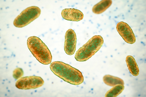
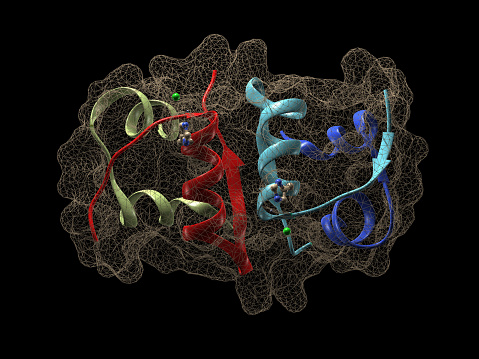
Free Images: "bestof:Galanin.png PSEUDOMONAS AERUGINOSA LECTIN II PA-IIL COMPLEXED WITH LACTO-N-NEO-FUCOPENTAOSE V LNPFV Jmol 11-17-10 Hawbash Hawramy/Jmol Development Team free"
Galanin.png
2CZP.pdb.png
3P1M.pdb1.png
1IP0.pdb.png
2W86 (Fibrillin).png
2VSZ.pdb1.png
3LL6.pdb1.png
Hexokinase.pdb1.jpg
Recoverin.png
Hexokinase.pdb2.jpg
Riboflavkinase.png
Neurcalcin.png
Miraculin.png
Cd59.png
Cd59b.png
Kynureninase2HZP.jpg
1O8R.pdb.png
1BQS.pdb.png
3MSC.png
PDB.png
Factor D.jpg
PDB1.png
Chimerinjmol.png
2BC4.pdb1.png
1IVA.pdb.png
3B7X.pdb.jpg
1GPS.jpg
3O5V.pdb.jpg
3LOY.pdb.png
3N7S.pdb.png
V receptor.png
Gprotein-coupled receptor kinase.png
Hexokinase 3O08.png
Jmol2WUX.pdb.jpg
Microcephalin.png
Project_Paperclip_Team_at_Fort_Bliss.jpg
Project_Paperclip_Team_at_Fort_Bliss_crop.jpg
Terms of Use   Search of the Day