Click Here for More Images from iStock- 15% off with coupon 15FREEIMAGES 
Leiden leaf c.s. under microscope
MRI scan of veins around the heart
Intestinal tract with holographic effect, 3d rendering. 3D illustration.
Mammogram Breast Examination
full abdomen ultrasound image aka diagnostic sonogram
Mammogram
Anatomy of the pelvis bones, including the ilium, ischium, sacrum, and pubis, photorealistic 3D illustration. Perfect for educational or medical purposes. Front view
Computational tomography image of the abdominal and pelvic regions.
MRI of the prostate gland, revealing an enlarged size, aids in diagnosing tumors, guiding treatment decisions, and monitoring prostate health.
Pancreatic neuroendocrine tumor, clear cell variant. Site: Pancreas.
Optical Coherence Tomography . ( OCT )
Close-up shooting of a green agate stone mineral surface isolated on white background. Its a macro shot shows nature beautiful details with back lit. Agate is a kind of natural gemstone. Abstract pattern. Good for Wallpaper texture background. Purple
ct scan step set of neck coronal view
skin science 3d
ultrasonography or ultrasound images of upper abdomen with image of gallbladder, liver, spleen, kidney, pancreas and aorta
Golden Turkish Lira Symbol
Wilms tumor, or nephroblastoma, light micrograph, photo under microscope
Carcinoid tumors are a type of slow-growing cancer that can arise in several places throughout your body. Carcinoid tumors, which are one subset of tumors called neuroendocrine tumors, usually begin in the digestive tract (stomach, appendix, small intestine, colon, rectum) or in the lungs. Site: Ovary
forming of underskin cellulite illustration
Computational tomography image of the abdominal and pelvic regions.
PAPS Smear study of a young women under microscopy showing Atrophic changes in uterus which is unusual at young age.
Breast MRI or magnetic resonance imaging of Breast both side with breast silicone implant in women for screening breast cancer.
Computed tomography of chest organs. CT scan. CT Abdomen Cross section View.
Showing Light micrograph Type of Tissue Human under the microscope in Lab.
Serous carcinoma is the most common type of ovarian cancer, accounting for approximately 75% of epithelial ovarian cancers.
Dicot plants stem show plant vascular tissue under light microscope view for botany education.
3D representation of the human body
Simple columnar epithelium  of an excretory duct of the pancreas. Light microscope picture.
CT angiography of lower limb..artery occlusive disease
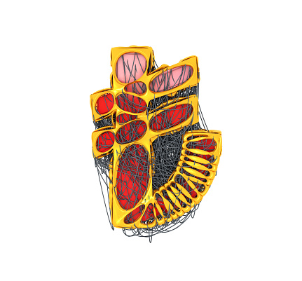
Free Images: "bestof:Gray911.png Summary with Anatomic Orientation This image taken from Gray's Anatomy is a view of the right-sided middle ear space with the external ear canal"
An_Odd_Planetary_Nebula_in_Hercules.jpg
NASA_Captures_"EPIC"_Earth_Image.png
Challenger.jpg
Naked human male body front anterior.png
Marley Beach Sand Dunes.jpg
STS-51-L_Accident.jpg
Hubble_Observes_Infant_Stars_in_Nearby_Galaxy_DVIDS727273.jpg
Naked human male body front anterior (annotated-id).png
Anatomía humana.png
PinkertonLincolnMcClernand.jpg
A-New-York-City-Fireman.jpg
F-18F_after_launch_from_USS_Abraham_Lincoln_(CVN-72).jpg
30_Doradus_in_the_Large_Magellanic_Cloud.jpg
A_Lucky_Observation_of_an_Enigmatic_Cloud.jpg
Probing_a_super-giant_shell_of_gas_and_stars.jpg
A_Vapour_of_Stars.jpg
Return_to_the_Carina_Nebula.jpg
Sarychev Volcano edit.jpg
A-New-York-City-fireman.jpg
HD40307.svg
GJ581orbits Vogt2010.svg
Flag of French language.png
Mellomore.svg
Araneae anatomic numbers.svg
Hand_external_anatomy,_dorsum.svg
Brachial plexus 2.svg
Brachial plexus 2 es.svg
Gray-backed_tern,_Pacific_Ocean,_April_1965..jpg
Image_of_antenna_support_for_LORAN_tower_on_Sand-Johnston_Island,_1963..jpg
Gray834 pl.svg
Compiled_image_of_aerial_photographs_taken_of_Sand-Johnston_island,_1964..jpg
Alice_Brown.jpg
Brain_diagram_ja.svg
Brain_diagram_pl.svg
Diagram of swine influenza symptoms EN.svg
ISS configuration 2009-07 cs.svg
Gray728.svg
Brain_diagram_without_text.svg
Jacqueline_L._Schroedter.jpg
Raven_Manet_E2_corrected.jpg
Gray696.svg
PinkLincMcC.jpg
Hans_Maler_-_Portrait_of_Wolfgang_Ronner_-_detail_letter.JPG
Circulatory System en edited.svg
Two views of cochlear mechanics (B).svg
Max_Valier_in_Rocket_Car_(1931).jpg
PD Diagram of swine flu symptoms EN.svg
left_to_right:_Clarence_Cook_Little_(1888-1971);_Edgar_Allen_(1892-1943);_Howard_Bancroft_Andervont_(1898-1981);_Madge_Thurlow_Macklin_(1893-1962);_Leiv_Kreyberg_(b._1896);_Gioacchino_Failla_(1891-1961);_and_Henri_Coutard_(1876-1950).jpg
Charles Leale - Brady-Handy.jpg
Circulatory System ru.svg
Construction_Progress_of_the_S-IC_Test_Stand-Block_House_Access_Tunnel.jpg
Gray911.png
Arteria_temporalis.png
Stages_in_the_development_of_the_external_sexual_organs_in_the_female_(from_Gray1119).png
Stages_in_the_development_of_the_external_sexual_organs_in_the_male_(from_Gray1119).png
Shaded_Relief_and_Radar_Image_with_Color_as_Height,_Bosporus_Strait_and_Istanbul,_Turkey_DVIDS712179.jpg
ISS015-E-15323 lrg.jpg
Image_from_page_554_of_"Local_and_regional_anesthesia_:_with_chapters_on_spinal,_epidural,_paravertebral,_and_parasacral_analgesia,_and_on_other_applications_of_local_and_regional_anesthesia_to_the_surgery_of_the_eye,_ear,_nose_and_throat,_and_to_dental_p.jpg
Dune Types in the Issaouane Erg Eastern Algeria.jpg
Orientation.PNG
External view of basal ganglia.jpg
Seattle, Washington, view taken from the Space Needle LCCN2010630406.tif.tiff
Seattle, Washington, view taken from the Space Needle LCCN2010630405.tif.tiff
Seattle, Washington, view taken from the Space Needle LCCN2010630407.tif.tiff
Seattle, Washington, view taken from the Space Needle LCCN2010630408.tif.tiff
Mammal middle ear.png
Gray1195_without_labels.png
Jimmy_Buffet_navy.jpg
Corps humain mâle nu devant (annoté fr).png
Gray927.png
Vulva-f.png
Bodleian Libraries, View of the canal in St. James's Park, the Queen's House, taken from the Parade.jpg
HumanEar.jpg
View of canal during sunset in Rawla.jpg
Gray1194.heb.PNG
Gray1211.heb.PNG
Gray1219.heb.PNG
Gray1215.heb.PNG
Gray1231.heb.PNG
Gray1232.heb.PNG
Gray235_he.png
Iliac_artery_bifurcation.PNG
Popliteal_artery.png
Axillary_space.png
Gray909-nolabels.png
Atlas_Collection_Image.jpg
Gray438he.png
Superior_thyroid.PNG
Iliac_artery_bifurcation_and_aorta.PNG
Anatomyofhumanbo1918gray_1280.jpg
Brachial plexus 2-2.png
Gray777.png
Gray827.png
Gray832.png
Gray918.png
Gray 111 - Vertebral column-coloured.png
Gray_188_-devided.png
Ascending_pharyngeal.PNG
Lingual_artery.PNG
Facial_artery.PNG
Terms of Use   Search of the Day