Click Here for More Images from iStock- 15% off with coupon 15FREEIMAGES 
Squamous cell carcinoma or squamous cell cancer (SCC or SqCC) is a cancer of a kind of epithelial cell, the squamous cell. These cells are the main part of the epidermis of the skin, and this cancer is one of the major forms of skin cancer. However, squamous cells also occur in the lining of the digestive tract, lungs, and other areas of the body, and SCC occurs as a form of cancer in diverse tissues, including the lips, mouth, esophagus, urinary bladder, prostate, lung, vagina, and cervix, among others. Despite sharing the name squamous cell carcinoma, the SCCs of different body sites can show tremendous differences in their presenting symptoms, natural history, prognosis, and response to treatment. Micrograph of squamous cell carcinoma of the head and neck
Kidney cancer: Clear cell carcinoma. fuhrman nuclear grade-II, Renal growth shows a malignant tumor, renal cell carcinoma in between renal tubules.
Disease X Cells. 3D Render
Bladder Cancer: High grade transitional cell carcinoma (TCC), Urothelial carcinoma
Backgrounds of Bacteria and Mold under the microscope in Lab.
CAT Scan lung lobe
Bronchitis and alveolitis in lung of horse infected with equine influenza virus, 200x magnification.
Digital Dispersed corona virus on petri dish. Animation of multiplication of CORONA VIRUS COVID-19
The uterine sarcoma is a malignant tumor that arises in the myometrium. The micrograph shows cancerous cells infiltrating the smooth muscle fiber fascicles of myometrium.
Serous carcinoma is the most common type of ovarian cancer, accounting for approximately 75% of epithelial ovarian cancers.
Cross section - Xylem is a type of tissue in vascular plants that transports water and some nutrients. Scientific research. Plant tissue Structure.
Closeup abstract color mixing of acrylic for use as background. Acrylic texture with marble pattern, blue marbling background
The study of plant tissues under the microscope in the laboratory.
Thyroid cancer: Microscopic image of Metastatic papillary carcinoma of thyroid. Reactive change. Lymph node carcinoma.
smoke lung with black deposit in microscopy
PAPS Smear study of a young women under microscopy showing Atrophic changes in uterus which is unusual at young age.
Mesenchymal dysplasia. Site: Placenta
Epidermis. Electron microscope micrograph showing a keratinocyte of spinous layer. The epithelial cell has a polygonal shape, central nucleus with nucleolus, cytoplasm full of keratin filament bundles, and numerous dark desmosomes crossing the intercellular spaces.
\
Host cells with spores (mold) are inside wood under the microscope for education.
Pneumonia coronavirus
Kidney cancer: Microscopic image of metastatic clear cell carcinoma of kidney, the most common type of renal cell carcinoma. Show brain tissue of malignant neoplasm, large atypical bizarre cells.
Silicosis lung with black deposit human pathology biosample under microscopy
An abstract shot of doctor's hands holding a corona virus cell and cut out lungs
Wilms tumor (also called Wilms' tumor or nephroblastoma) is a type of childhood cancer that starts in the kidneys. It is the most common type of kidney cancer in children.
Mucosa of a human stomach showing: surface epithelium, which invaginates to form the gastric pits. The fundic glands open into the pits and have parietal (pink) and chief (blue) cells.
\
Lung and Pituitary gland Human under the microscope in Lab.
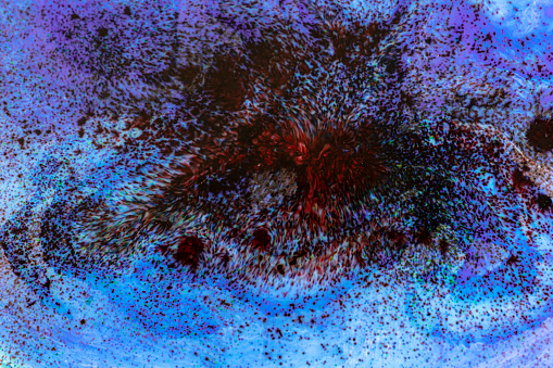
Free Images: "bestof:Human Lung Cells (6680207077).jpg Photomicrograph showing human lung cells infected with cytomegalovirus Cell at center clearly shows virus particles large dark"
Ebola_virus_particles_found_both_as_extracellular_particles_and_budding_particles_from_chronically-infected_African_green_monkey_kidney_cells_..jpg
Humanstemcell.JPG
blood-cells-cells-human-543484.jpg
3._Red_blood_cells_showing_signs_of_yeast.jpg
cancer-cells-cells-scan-541954.jpg
Ebola_Virus_Particles.jpg
anatomy-man-human-face-body-lips-254129.jpg
anatomy-woman-human-face-body-254120.jpg
woman-human-pair-man-face-body-254133.jpg
Ebola_Virus_Particles.jpg
Secondary tumor deposits in the liver from a primary cancer of the pancreas.jpg
lava-lava-rock-lava-fields-boulders-571799.jpg
annecy-france-channel-water-1687832.jpg
virus-infected-cells-dna-disease-213708.jpg
Riassorbimento bicarbonati e respirazione cellulare.svg
Ebola Pathenogensis.svg
Ebola Pathenogensis path.svg
Ebola Pathenogensis path-hu.svg
Complete neuron cell diagram numbered large numbers.svg
portugues-sexo_alemão-sex_ingles-sex_africano-seks_russo-секс_albanes-sex_chines-性別_bulgaro-секс_croata-spol_gales-rhyw_coreano-섹스_frances-sexe_japones-セックス_filandes-sukupuolesta_arabe-الجنس_prontos_ai_tens_em_15_linguas_a_p.jpg
Metastasis sites for common cancers.svg
Complete neuron cell diagram en.svg
Complete neuron cell diagram it.svg
Energy change inventory, 1971-2010.svg
Diagram human cell nucleus.svg
Diagram human cell nucleus pl.svg
Diagram human cell nucleus fr.svg
Diagram human cell nucleus es.svg
Health benefits of apples.svg
Stem cell treatments.svg
Complete neuron cell diagram without text.svg
Complete neuron cell diagram zh-hant.svg
Complete neuron cell diagram pl.svg
Complete neuron cell diagram numbered.svg
Complete neuron cell diagram es.svg
Complete neuron cell diagram zh.svg
Complete neuron cell diagram pt.svg
Cellular tight junction-en.svg
Cellular tight junction-fr.svg
Animal mitochondrion diagram en (edit).svg
Complete neuron cell diagram ru.svg
Thyroid hormone synthesis.svg
Adherens Junctions structural proteins-fr.svg
Adherens Junctions structural proteins.svg
immune-system-blood-cells-virus-1132182.jpg
lungs-diagram-anatomy-body-human-41562.svg
lungs-human-body-anatomy-body-311005.svg
t-helper-cell-cell-immune-system-1123292.png
Mitosis cells sequence.svg
human-digestive-system-163714.jpg
man-woman-schema-body-anatomy-144378.jpg
Hand_holding_a_cigarette.jpg
petri-dish-bacteria-laboratory-311960.svg
Pulmonary tuberculosis symptoms.svg
T_cell_activation.svg
T_cell_activation_-_ru.svg
cancer-smoke-healthcare-death-1088631.jpg
Cellular tight junction keys.svg
B_cell_activation.svg
bacteria-macro-art-abstract-1527639.jpg
F4 roots by 24-cell duals.svg
Zinc-carbon-numbered.svg
Cellular tight junction-sv.svg
Cellular tight junction-it.svg
Mouvement diaphragme respiration.svg
B_cell_activation_-_ru.svg
man-boy-stylish-web-worldwide-www-475560.jpg
man-boy-stylish-internet-network-475559.jpg
woman-stylish-at-internet-network-163426.jpg
man-face-at-internet-network-694536.jpg
Smallpox versus chickenpox english plain.svg
Scheme simple diffusion in cell membrane-zh (zh-cn).svg
Scheme simple diffusion in cell membrane-zh (zh-tw).svg
Scheme simple diffusion in cell membrane-en.svg
Scheme simple diffusion in cell membrane-pl.svg
Animal mitochondrion diagram unlabelled.svg
woman-stylish-at-internet-network-569563.jpg
man-boy-stylish-internet-network-475561.jpg
man-boy-stylish-internet-network-475558.jpg
man-boy-stylish-internet-network-475557.jpg
digital-zeros-ones-woman-stylish-388075.jpg
man-boy-stylish-internet-network-516336.jpg
man-old-senior-stylish-at-1660853.jpg
man-stylish-at-internet-network-1657004.jpg
Scheme simple diffusion in cell membrane-es.svg
Scheme simple diffusion in cell membrane-de.svg
Scheme simple diffusion in cell membrane-fr.svg
Gray5.svg
Gray5-ja.svg
Illu capillary numbers.svg
Gray72-en.svg
Outer hair cell and Deiter's cell.svg
Smallpox versus chickenpox neutral plain.svg
Smallpox versus chickenpox latin plain.svg
Vaiolo VS Varicella.svg
Gray72-de.svg
Alveolus diagram.svg
Gray72-Human Bone marrow-Medula osea-es.svg
Terms of Use   Search of the Day