Click Here for More Images from iStock- 15% off with coupon 15FREEIMAGES 
3D Illustration Concept of Human Circulatory System Heart Anatomy
Front view of woman's thigh and knee muscles with names
A lovely cheerful six year old boy smiling with broken incisors and half grown incisor teeth with Iceland  flag painted on his face.  Black background. Black hair falling on his forehead. Black eyes. white teeth. Face along with lips and ears is also painted
Two anterior or front views of the human male body and skeleton xray 3D rendering illustration on blue background. Anatomy, medical, skeletal or bone system, science, biology, osteology concepts.
Glutenous Maximums: This is the largest and most superficial of the gluten muscles. It is responsible for the extension and lateral rotation of the hip joint. The glutenous maximums is what gives the buttocks its shape and provides power during activities like running, climbing, and jumping.
Human anatomy model. Photography in high resolution.Similar pictures from my portfolio:
The vertebral column surrounds the spinal cord which travels within the spinal canal, formed from a central hole within each vertebra
The hud interface of the futuristic medical program is a control panel with digital three-dimensional MRI visualization, which allows you to study the condition of joints, spine, muscles and soft tissues in detail.
3D Illustration Concept of Human Circulatory System Heart Anatomy
MRI scanning of the human body skeletal, muscular and nervous systems with abdomen region view. / You can see the animation movie of this image from my iStock video portfolio. Video number:
Human anatomy detail of shoulder. Muscle, bone structure, arteries. On plain studio background. Professional lighting.
3D Render Illustration of a man clutching his chest in pain.
Muscle maps of an adult female, anatomy views of the front, back and side and the corresponding nude body, isolated on white, 3d render
Medical illustration about Sacrum.3d rendering. No AI create.
Labeled human anatomy Erector Spinae back muscles in isolation on the skeletal system from a posterior view on a white background.  3D rendering.
3D illustration of female muscular system of an exercising person.
X-ray look of the human heart and vascular system
Xray lateral or profile view of the laryngopharynx or hypopharynx 3D rendering illustration with male body contours. Human anatomy, medical, biology, science, medicine, healthcare concepts.
3D Illustration Concept of Human Muscular System Deltoideus, Triceps and Latissimus Dorsi Muscles Anatomy
X-ray machine/medical digital X-ray machine is needed to photograph the patient's body from all sides, as well as to take X-rays of internal organs and the skeleton.
The chest muscles, specifically the pectoralis major and pectoralis minor, are important muscles located in the anterior (front) part of the chest. They play a significant role in various upper body movements, particularly those involving the shoulder and arm.
Vitruvian man. Woman also available.
Man's upper body muscles frontal anterior view labeled on a white background.
Red map pointers with flag USA and United Kingdom isolated on white background. Map pin icon. GPS place marker. Navigation and travel location. Start and end trip symbol.
Close-up of the female pelvis in the hands of an athletic, healthy woman. Gynecology, women's healthcare and medicine and healthy body concept.
skin science 3d
Woman anatomy cardiovascular system with skeleton and internal organs. Ghost effect on white background.
France flag textile cloth fabric texture
3d rendered illustration of the female anatomy
Remember and honor American culture digital concept
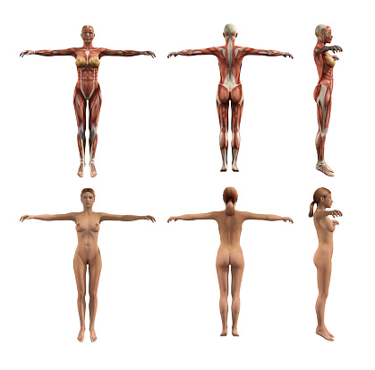
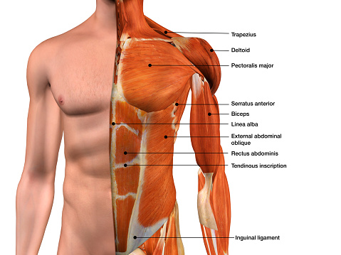
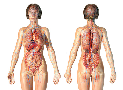
Free Images: "bestof:Human anatomy ru 2.jpg en Human male and female - anatomical features pointed out ru Анатомия человека По с� авнению с"
Terms of Use   Search of the Day