Click Here for More Images from iStock- 15% off with coupon 15FREEIMAGES 
Accurate xray image of human skeletal system with adult male skeleton and body contours on blue background 3D rendering illustration. Anatomy, medical, healthcare, science, osteology, concept.
Xray view of a patient with an injured spine and suffering. / You can see the animation movie of this image from my iStock video portfolio. Video number: 1453180480
Anatomy of the pelvis bones, including the ilium, ischium, sacrum, and pubis, photorealistic 3D illustration. Front view. Perfect for educational or medical purposes.
X-ray of skeleton
The sacrotuberous ligament (STL) is a stabiliser of the sacroiliac joint and connects the bony pelvis to the vertebral column 3d illustration
\
3D Illustration Concept of Human Urinary System Kidneys with Bladder Anatomy
Magnetic resonance images of the brain (MRI brain). Computed resonance imaging of the human brain.
Front and back views of full human skeleton with male body 3D rendering illustration isolated on black. Anatomy or medical diagram with each bone or group of bones labeled with colors.
3D illustration of male human body skeletal system.
Male Skeleton in full body front view with labeled bones on a white background, computer generated image.
Human brain MRI and AI technology.
Infographic diagram of lower half human skeleton anatomy system anterior view- 3D- Human Anatomy- Medical Diagram- educational and Human Body concept- Isolated on white background
Human skeleton for demonstration made of plastic
Magnetic resonance image of human spine with lordosis, disc herniation and spondylolisthesis l5-s1
X-ray of the human mouth showing dental
Anterior or front view of accurate human skeletal system with skeleton bones of adult male isolated on white background 3D rendering illustration. Anatomy, medical, science, osteology concept.
Backlit image of fake bones made of resin.
The lumbar vertebrae are, in human anatomy, the five vertebrae between the rib cage and the pelvis .
CAT scan of human abdomen...man with aneurysm of abdominal aorta
3D illustration of sanitary piping
Xray of ones manhood.
CT (Computed Tomography) scan of Whole abdomen. Soft tissue mass in epigastric region, lymphadenopathy, GIST, hugely distended stomach containing food residue.
model of the human spine isolated on white
Femur bones in red color with body 3D rendering illustration isolated on white with copy space. Human skeleton and leg anatomy, medical diagram, osteology, skeletal system, science concepts.
Human brain MRI.
A triangular bone in the lower back formed from fused vertebrae and situated between the two hip bones of the pelvis.
The intertransverse ligaments comprise sheets of connective tissue that extend from one transverse process to the next 3d illustration
Computational tomography image of the abdominal and pelvic regions.
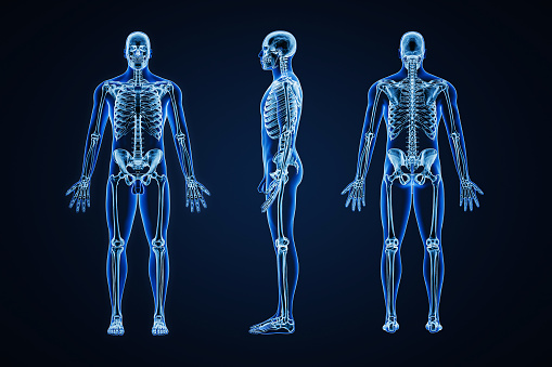
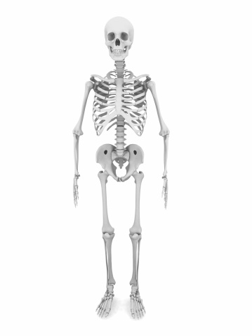
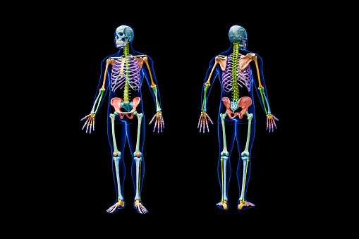
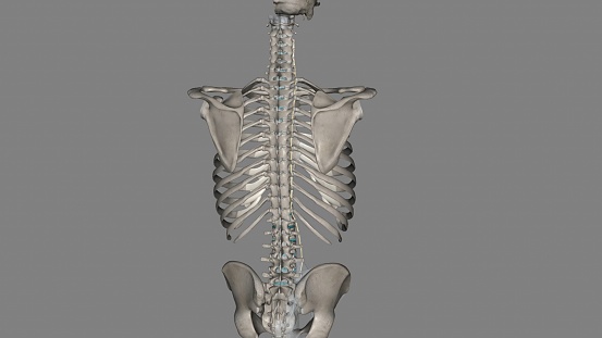
Free Images: "bestof:Human skeleton front ba.svg ba Кеше һөлдәһе алдан кү� енеш ҡыҙыл һыҙыҡта� айы� ым һөйәктә� ҙе кү� һәтә"
Human skeleton front ba.svg
Human skeleton back ba.svg
George_Stubbs_-_A_Comparative_Anatomical_Exposition_of_the_Structure_of_the_Human_Body_with_that_of_a_Tiger_and_a_Co..._-_Google_Art_Project_(2349603).jpg
Terms of Use   Search of the Day