Click Here for More Images from iStock- 15% off with coupon 15FREEIMAGES 
Viral infection disease attacking cell abstract idea illustration.
COVID-19 new mutation variant - 3d rendered image. Pandemic wave.\nViral Infection concept. Delta, Omicron (B.1.1.529), Centaurus (B.1.1.529.2.75) (BA.2.75), Aeterna (BA.4.6) variant. Sars-cov-2, 2019-nCoV, Coronavirus.\nAntibody, Antigen, stealth, Vaccine technology concept.\nB117 - COVID-19 Variant,\nSARS-CoV-2 BA.2 Variant,\nSARS-CoV-2 BA.5 Variant,\nSARS-CoV-2 Omicron Variant,\nSaRS-CoV-2 Deltacron Variant
Illustration of Respiratory Syncytial Virus or RSV
Four COVID-19 viruses in diffrent colors isolated on white background.
orange: RNA primer; red: RNA template; light blue: proofreading riboexonuclease; mid blue: helicase; dark blue: RNA directed RNA polymerase; violet: non structural proteins (NSPs).
Inside protein molecule
Human adenovirus model. Structure of the adenoviridae virion: penton base, nucleocapsid with DNA genome, capsomers, proteins. Acute respiratory viral infections, ARVI disease, 3D medical science mixed media illustration
Virus Cells of the Adenovirus Family in Icosahedral Shape
3d rendering of IgA molecules neutralize or block the activity of virus and prevent their attachment to host cells
COVID-19 new mutation variant Pirola (BA.2.86) - 3d rendered image. Pandemic wave.\nViral Infection concept. Concept image for Delta, Omicron (B.1.1.529), Centaurus (B.1.1.529.2.75) (BA.2.75), Aeterna (BA.4.6) variant. Sars-cov-2, 2019-nCoV, Coronavirus.\nAntibody, Antigen, stealth, Vaccine technology concept.\nB117 - COVID-19 Variant,\nSARS-CoV-2 BA.2 Variant,\nSARS-CoV-2 BA.5 Variant,\nSARS-CoV-2 Omicron Variant,\nSaRS-CoV-2 Deltacron Variant
The coronavirus spike protein that mediates coronavirus entry into host cell
A group of viruses together as seen under a microscope
Background with viruses. Adeno-associated virus serotype 1. Virus is used for gene therapy. 3D illustration
SARS-CoV-2 XBB 1.5 Variant. Kraken. Coronavirus. 3D Render
Monkeypox virus is a smallpox-like viral infection transmitted from animals to humans. 3D illustration
Digitally painted on Tablet, Pixelated Planets, Abstract Background
Virus variant, coronavirus, spike protein. Omicron. Covid-19 seen under the microscope. SARS-CoV-2, 3d rendering
Endosomes - 3d rendered image of Endosomes are membrane-bound organelles. They are parts of endocytic membrane transport pathway.  Endomembrane system in cells. Bilayer Lipid Membrane. Abstract medical illustration. Microscopic view. Microbiologic research concept.
Subvariant Virus Spreading as viral pathogens Mutating variants and mutation as a transmissible health risk concept and new COVID-19 outbreak or coronavirus mutations and influenza background as a 3D render.
Concept of COVID-19 cells, with blue background, and medicine pills around. Focus with depth of field. Cure concept render.
Nucleosome, molecular model. 3D illustration. The nucleosome consists of a DNA double helix wrapped around a core of histone proteins.
Digitally generated illustration of a SARS-CoV-2 virion on black background.\n\nThe scene was rendered with photorealistic shaders and lighting in Autodesk® 3ds Max 2020 with V-Ray Next with some post-production added.
3D render of an abstract medical background with virus cells
Realistic model of A/H1N1 virus on the basis of photos from electronic microscope.
Biological protein and molecule, 3d rendering. 3D illustration.
Bacteria pathogen - 3d rendered microbiology image. Illustrate of Lactobacillus Bulgaricus Bacteria, Microbacterium Tuberculosis Bacteria (MTB). Medical research, health-care concept. SEM (TEM) scanning view
Feline panleukopenia virus, also called as feline infectious enteritis virus or cat plague. 3D illustration
Texture pattern on marble of stone surface of agate slice.
The NEK-7 independent NLRP3 inflammasome in it's hexameric conformation with ADP (red) bound. Inflammasomes are a promising target for the treatment of autoimmune diseases. PDB entry: 7zgu.
Futuristic technology
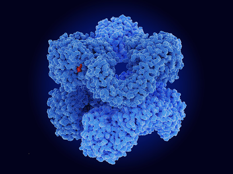
Free Images: "bestof:Jmol2WUX.pdb.jpg Polyhedrin protein 2WUX from virus baculovirus Autographa californica see https //en wikipedia org/wiki/Baculoviridae Structure_of_the_virion"
Hepatitis B virus v2.svg
Jmol2WUX.pdb.jpg
Computer-generated image of the structure for C-reactive protein.jpg
PBB Protein AKR1A1 image.jpg
Denise Ney, PhD, RD.jpg
Biochemical Protein Purification Challenge.jpg
2014 Ebola virus epidemic in West Africa.png
Terms of Use   Search of the Day