Click Here for More Images from iStock- 15% off with coupon 15FREEIMAGES 
Creative paper concept. Different types of ovarian cysts. Yellow cyst, follicular cyst, endometrioid cyst, dermoid cyst
Rosai–Dorfman disease (RDD) is a rare proliferative histiocytic disorder of unknown etiology. RDD typically presents with generalized lymphadenopathy and polymorphic histiocytic infiltration of the lymph node sinuses; however, occurrences of extranodal soft tissue RDD may rarely occur when masquerading as a soft tissue sarcoma.
Uterus or uterine cancer. Medical concept as cancerous cells spreading in a female reproductive system. 3d illustration
Radiology report. On the right, lateral breast there are two intramammary lymph nodes with fatty hilum measuring 0.5 and 0.6 cm respectively. No suspicious breast mass or axillary lymphadenopathy. Moderately dense breasts with 50 to 75 % glandular tissue.  Bi-RADS 2 : benign findings.
PAPS Smear study of a young women under microscopy showing Atrophic changes in uterus which is unusual at young age.
Full size digital screening mammogram of a new breast tumor in the retroareolar region of the right breast causing nipple retraction. An ultrasound guided biopsy was performed diagnosing invasive ductal carcinoma. This is the most common type of breast cancer, accounting for nearly 80% of cases. Breast cancer treatment has come a long way in the last few decades, seeing more funding than any other human disease. The first step to a cure is catching it early with screening mammograms such as this case so that the tumor can be removed before it spreads (metastasizes) out of the breast.
Breast Cross Section of Fat .
Doctor holding and showing  human Inner ear model of auditory system
skin detail
Focal nodular hyperplasia (FNH) is a benign liver lesion that is composed of a proliferation of hyperplastic hepatocytes surrounding a central stellate scar. Typically, FNH is a solitary lesion that is more commonly seen in women.
prostate cancer, prostatic hyperplasia, Structure of male prostate, cross section, 3d illustration
Computational tomography image of the abdominal and pelvic regions.
Skin tumors, moles and spots, 3d section of the skin layer. Three types of skin cancer, squamous cell carcinoma, basal cell carcinoma, melanoma. 3d render
Ultrasound monitor
Bacterial vaginosis is a genital infection caused by bacteria, mainly Gardnerella Vaginalis. 3D illustration
Image taken with Flir T420 infra red digital camera. Each color represents different temperatures, as is shown on spectrum scale on right side of image.
Nine images of fetus ultrasonography.
A meningioma is a tumor that arises from the meninges — the membranes that surround the brain and spinal cord. Although not technically a brain tumor, it is included in this category because it may compress or squeeze the adjacent brain, nerves and vessels.
The xylem and phloem that make up the vascular tissue of the stem are arranged in distinct strands called vascular bundles, which run up and down the length of the stem
Four, side view, X-ray pictures of a woman's two breasts. On the right, lateral breast there are two intramammary lymph nodes with fatty hilum measuring 0.5 and 0.6 cm respectively. No suspicious breast mass or auxiliary lymphadenopathy. Moderately dense breasts with 50 to 75 % glandular tissue.  Bi-RADS 2 : benign findings.
Stomach cancer, symptoms, stages, treatment, 3d illustration
\
Hyaline cartilage, Elastic cartilage and Bone Human under the microscope in Lab.
Abstract technological design of an artificial molecular structure in many colors. Horizontal wide composition.
Female uterus with virus bacteria. 3d illustra
Squamous cell carcinoma of the lung is a type of lung cancer. It occurs when abnormal lung cells multiply out of control and form a tumor.
Infographic explaining depth of field and the corresponding aperture values with their effect on blur and light
3d render, abstract background with rock
3D cartoon and Gaussian surface models, PDB 6gu2, white background
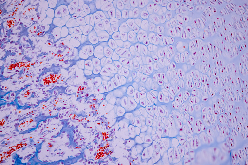
Free Images: "bestof:LH and FSH in menstrual cycle.svg Reference ranges for luteinizing hormone and follicle-stimulating hormone throughout the menstrual cycle Detail from picture"
An_Odd_Planetary_Nebula_in_Hercules.jpg
LH and FSH in menstrual cycle.svg
Estradiol and progesterone in menstrual cycle.svg
Hormones estradiol, progesterone, LH and FSH during menstrual cycle.svg
Hormones estradiol, progesterone, LH and FSH during menstrual cycle pt.svg
Hormones estradiol, progesterone, LH and FSH during menstrual cycle es.svg
Hormones estradiol, progestérone, LH et FSH pendant cycle menstruel.svg
Peschls three loops extended to social construction.svg
Hormones in menstrual cycle.png
Hormones estradiol, progesterone, LH and FSH during menstrual cycle.png
Luteinizing hormone (LH) during menstrual cycle.png
Follicle-stimulating hormone (FSH) during menstrual cycle.png
Luteiniseeriv hormoon (LH) menstruaal tsyĂĽkli ajal.png
Progesterone during menstrual cycle.png
Estradiol during menstrual cycle.png
Progesterone-3D-spacefill.png
Progesterone-3D-balls.png
Progesterone-3D-skeletal-sticks.png
Lining_of_Uterine_Wall.jpg
FSHA+B+receptor 1XWD.png
Melatonin molecule ball.png
Melatonin molecule spacefill.png
Biketrail map1.jpg
CloudSat-CALIPSO_Launch_Successfully,_Image_of_the_Day_DVIDS749611.jpg
Terms of Use   Search of the Day