Click Here for More Images from iStock- 15% off with coupon 15FREEIMAGES 
Computer generated 3D illustration with the dinosaur Argentinosaurus
plant Zea Stem C.S. under light mircoscope with white background
Lambeosaurus dinosaur walking isolated in white background - 3D render
Carp tail close-up isolated on white background
Epidermis. Electron microscope micrograph showing a keratinocyte of spinous layer. The epithelial cell has a polygonal shape, central nucleus with nucleolus, cytoplasm full of keratin filament bundles, and numerous dark desmosomes crossing the intercellular spaces.
Photomicrograph of algae, Cosmarium turpinii, a desmid. Live specimen. Wet mount, 40X objective, transmitted brightfield illumination.
A collection of animals, fish, insects that lived during the Carboniferous Period.
Swimming slowly, a large white sturgeon and salmon cruise the water in the Bonneville Fish Hatchery and Sturgeon Center along Oregon’s Columbia River.
Catfish isolated on white background. Clipping path inside.
Trematode parasite under a microscope
Parasaurolophus dinosaur walking next to gingko trees and onychiopsis plants - 3D render
Liver Fluke Section under light microscope with white background
Indian pike conger isolated on white, congresox talabonoides
Nature Photos of the “flat” top of  Table Mountain - Cape Town, Western Cape
Planarian parasite (flatworm) under microscope view.
Hippopotamus
Computer generated 3D illustration with adult Paraceratherium and baby Paraceratherium
Female manatee and calf near Titusville, Florida
Carnivorous dinosaurs set isolated in white background - 3D render
Cheyletiella blakei Mite - Small Cat pest view under a microscope
A toy dinosaur.
Photomicrograph of filamentous blue-green algae, Oscillatoria species. Each band along the stand is one cell. This filament is very short probably having recently parted from a longer one. Filaments are typically many times longer. This algae is mobile, it moves and twists through the water. San Francisco Bay, California, USA. Live specimen. Wet mount, 40X objective, transmitted brightfield illumination. Note - motion blur of live specimen, very shallow depth of field, chromatic aberration and uneven focus are inherent in light microscopy.
A close up view of a lowland Tapir outdoors in a zoo enclosure.
Nature Photos of the “flat” top of  Table Mountain - Cape Town, Western Cape
Cymbospondylus was an Ichthyosaur carnivore that lived in the seas of Germany, and Nevada during the Triassic Period.
Pigmy hippo in white background
Deinocheirus dinosaur walking isolated in white background - 3D render
Swimming Asian water monitor in Lumphini Park, which is a large public park in the center of Bangkok the capital of Thailand
Photograph of autotrophic Euglena through microscope under brightfield illumination.
Hippopotamus (Hippopotamus amphibius) eats hay
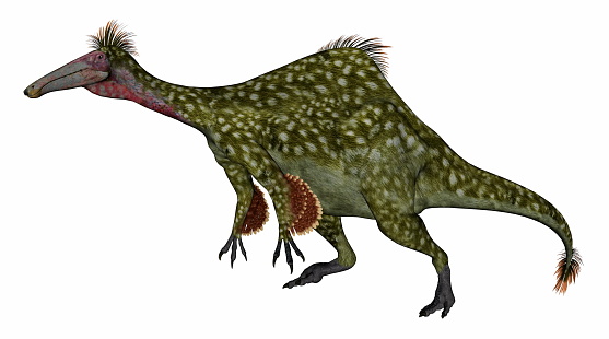
Free Images: "bestof:Limusaurus forelimb 2.png en Diagram of the forelimb of the herbivorous ceratosaur Limusaurus inextricabilis showing characteristic mesomelia and hypophalangia"
Limusaurus-arm.png
Limusaurus forelimb 2.png
Gene expression bat wing.png
Brachiosaurus-nougaredi.png
Bird and dino hand difference debate.JPG
Terms of Use   Search of the Day