Click Here for More Images from iStock- 15% off with coupon 15FREEIMAGES 
Striped mosquitoes life circle with white background. Life cycle of mosquitos isolate on white background.
Mosquito in Kruger NP
Mosquito Borne Virus risk like Zika  Dengue and Malaria spread through Aedes and Anopheles mosquitoes causing infectious diseases with 3D illustration elements.
Mobile shot
Bacteria Aggregatibacter, 3D illustration. Aggregatibacter aphrophilus and A. actinomycetemcomitans, Gram-negative bacteria, part of the normal flora of the mouth and throat, also cause endocarditis
Photomicrograph of filamentous blue-green algae, Oscillatoria species. Each band along the stand is one cell. This filament is very short probably having recently parted from a longer one. Filaments are typically many times longer. This algae is mobile, it moves and twists through the water. San Francisco Bay, California, USA. Live specimen. Wet mount, 40X objective, transmitted brightfield illumination. Note - motion blur of live specimen, very shallow depth of field, chromatic aberration and uneven focus are inherent in light microscopy.
Cotton whitefly (Bemisia tabaci) adults, eggs and larvae on a leaf underside.
plant Zea Stem C.S. under light mircoscope with white background
Close up a Mosquito sucking human blood_set B-4
Extreme close-up shot of the mosquito sucking blood on the male hand
Global Mosquito Disease spread as mosquito-borne illness as climate change concept with mosquitoes resulting in West Nile Virus Zika or malaria.
Fly on a apple spitting out waterbubbles, orange background, bokeh,
gnats; mosquitoes; larvae
West Nile are invading blood system in blood vessels. Easy to crop for all your social media and print sizes.
Dermatophyte fungi, 3D illustration. Microsporum, Trichophyton, and Epidermophyton, the causative agents of ringworm, tinea, skin, hair and nail disease
Jungle Mosquito Stock Photo
Mosquito
3d rendering of Easter eggs background
Vector Borne Disease as pathogens and parasites transmitted by vectors as ticks a fly or mosquito and fleas as a flea infection or spreading a virus through mosquitos.
Pholcus phalangioides Cosmopolitan Cellar Spider. Digitally Enhanced Photograph.
This is an illustration of the life cycle of a carrion fly including the maggots, emerging pupa and adult fly.
Common house fly close up CGIs, alpha result differs between white and black backgrounds
It looks like a macro of mosquito insects that have become mummified because they have been entangled for a long time by the sticky liquid from Pinguicula
Simuliidae, black fly (Diptera) in Baltic amber, Eocene, approximately 56 - 34 million years ago. Image taken with extreme macro and focus stacking technique.
Mosquito under microscope, selective focus, light micrograph
Extreme Close-up of Housefly on Mosquito Net.
In eukaryotic cells, DNA polymerase beta (POLB) performs base excision repair (BER) required for DNA maintenance, replication, recombination, and drug resistance. 3D cartoon model in two perpendicular projections, white background
Spider and its prey isolated on white. Studio shot.
Microscopic of paramecium and amoeba
Bright green fly insect
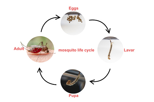
Free Images: "bestof:Malaria LifeCycle(French version).GIF Malaria Plasmodium Plasmodium vivax Plasmodium ovale Plasmodium malariae Plasmodium knowlesi malaria_LifeCycle"
female-mosquito-anopheles-insect-1332276.jpg
Malaria_LifeCycle(French_version).GIF
The spatial distribution of Plasmodium vivax malaria endemicity map in 2010 globally.png
"Life_History_of_the_Malaria_Parasite_(Plasmodium_Vivax)_in_man_and_the_Anopheles_Mosquito"_-_NARA_-_514892.tif
"Life_History_of_the_Malaria_Parasite_(Plasmodium_Vivax)_in_man_and_the_Anopheles_Mosquito"_-_NARA_-_514892.jpg
Microgametocyte_of_the_parasite_Plasmodium_vivax_PHIL_2727_lores.jpg
Plasmodium kierto.png
Immature_and_mature_trophozoites_of_the_Plasmodium_vivax_parasite_PHIL_2720_lores.jpg
Malaria_parasite_life_cycle-NIAID.jpg
Malaria_lifecycle-CDC.gif
Birds-eye trophozoite of P. malariae in a thin blood smear.jpg
Schaudinn_1902.JPG
P_vivax_trophozoite4.jpg
Malaria_Plasmodium_gallinaceum.jpg
Plasmodium falciparum (malaria) parasite in blood.jpg
Plasmodium_falciparum_(Estivo-Autumnal_Malaria)_Giemsa_stain.jpg
Plasmodium_life.jpg
Plasmodium_falciparum_nephrosis_edema_PHIL_3894_lores.jpg
Plasmodium_falciparum_rings_form_parasites4885_lores.jpg
Plasmodium.jpg
Hemoglobinopathies and the pathogenesis of Plasmodium falciparum malaria - Journal.ppat.1003327.g001.png
Gametocytes_plasmodium_5952_lores.jpg
Hemoglobinopathies-Slicing-the-Gordian-Knot-of-Plasmodium-falciparum-Malaria-Pathogenesis-ppat.1003327.g001.jpg
Macrogametocyte_of_the_parasite_Plasmodium_falciparum_PHIL_2701_lores.jpg
PLDH Malaria Antibodies.jpg
Hemozoin crystals.jpg
Glucose-6-Phosphate Dehydrogenase activity stain.jpg
Falciparum_ring.jpg
MalariacycleBig.jpg
TDR Plasmodium Step1.JPG
TDR Plasmodium Step3.JPG
TDR Plasmodium End.JPG
Plasmodium zyklus.png
Plasmodium falciparum 02.jpg
Plasmodium falciparum in Red Blood Cells.jpg
Plasmodium glutamate dehydrogenase precipitation.jpg
Ghana2.JPG
Plasmasexlifec.jpg
P.falciparum schizont.jpg
Magnetic collection.jpg
P.falciparum Gelatine enrichment.jpg
Birefringent P.falciparum Pigment.jpg
LDH.PNG
NordGhana.JPG
Blood stages of P. falciparum.jpg
Synchronsed P falciparum cultures.jpg
Tropeninstitut berlin.jpg
P.falciparum hemozoin.jpg
Blood stages of P. falciparum over 48h.jpg
A_manual_of_poisonous_plants_(Page_159)_(6005305428).jpg
Plasmepsin.png
Gradient centrifugation of infected Red Blood Cells.jpg
Haeckel_Mycetozoa.jpg
Terms of Use   Search of the Day