Click Here for More Images from iStock- 15% off with coupon 15FREEIMAGES 
Microscopic photo of a professionally prepared slide demonstrating Plasma cell myeloma from bone marrow aspirate. Wright Giemsa stain.
Epidermis. Electron microscope micrograph showing a keratinocyte of spinous layer. The epithelial cell has a polygonal shape, central nucleus with nucleolus, cytoplasm full of keratin filament bundles, and numerous dark desmosomes crossing the intercellular spaces.
Plant tissue Structure, section (tissue) of stem plant tissue under a light microscope.
3d cell organelle - nucleus (Chromatin,  Rough Endoplasmic Reticulum, Nucleus Shell, Nucleoplasm, Nucleolus)
The Raf/MEK/ERK (MAPK) propagates growth signals in the cell. Misregulation can lead to cancer.\nThe red dots on the active EGFR are phosphorylated tyrosines. PDB entries: 1egf, 1nql, 1m17, 2jwa, 3njp, 2gs6, 1gri, 1xd2, 3ksy, 5p21, 6xi7, 6q0j, 2y4i,
Squash preparation of  onion root tip cells stained to reveal chromosomes,  Bright field illumination, light microscopy.  This image was taken with the X40 objective lens.  Depth of focus is extremely low at this magnification.  Smaller apertures give greater depth of focus, but image quality deteriorates and resolution is greatly reduced.  The aperture in this image was optimised for contrast and resolution. Chromatic abberation is inevitable in light microscopy, but has been kept to a minimum in this image.
Senescent Cell as zombie cells as a symbol for Cellular Senescence and aging in living tissue as secreted molecules from the cell may repair tissues as age related changes as a positive or negative effect on health.
Abstract blue cell pattern, spots, cool, virus, background effect, texture
Internal structure of an animal cell, 3d rendering. Section view. Computer digital drawing.
Hashimoto thyroiditis is an autoimmune disorder involving chronic inflammation of the thyroid.
Motor Neuron cells under the microscope
plant Zea Stem C.S. under light mircoscope with white background
3D illustration of a cluster of Fat cells isolated on white.
Particles, medical concept
Human large intestine tissue under microscope view. Histological for human physiology.
Human embryonic stem cells
Mitosis process, division of cell. Isolated on black background. 3d render
Mesenchymal dysplasia. Site: Placenta
Backgrounds of Bacteria and Mold under the microscope in Lab.
Genetics background. 3D render.
Illustration of animal cell anatomy
Photomicrograph of
3d rendering of Human cell or Embryonic stem cell microscope background.
Human Cell Division. Early Stage Embryo. Stem Cell Research
New Cancer treatment with vibrating molecules breaking the membranes of malignant cells as an oncology therapy fighting Cancerous Growth and Metastasis anatomy concept as a molecular jackhammer effect.
Pancreatic neuroendocrine tumors (NETs), or islet cell tumors, are a type of cancer that starts in the pancreas.
Cells isolated on white background
plant rape flower cross section under light mircoscope with white background
Microscopic image of onion root tip cells undergoing mitosis. Anaphases and metaphases. 1000x magnified
Curly marble stone with fashion veins.
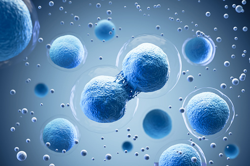
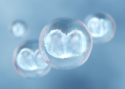
Free Images: "bestof:Mitosis cells sequence.svg A series of cell diagrams showing the mitosis division of eukaryotic cells Own work The cells are extracted from <gallery> Image"
Tissue Engineering; Biomaterials for the Heart (5881107872).jpg
paprika-fruit-bowl-chromoplasts-1582256.jpg
Nude mouse (1).jpg
Project_365_#287:_141009_A_Battery_of_Batteries.jpg
jail-cells-jail-penitentiary-police-429638.jpg
3._Red_blood_cells_showing_signs_of_yeast.jpg
Humanstemcell.JPG
agar-breeding-ground-red-60571.jpg
oil-glands-loosestrife-1582254.jpg
jail-prison-police-crime-law-429633.jpg
jail-prisoner-captive-police-crime-429639.jpg
microscope-sea-urchin-egg-splitting-1276131.jpg
Ebola_virus_particles_found_both_as_extracellular_particles_and_budding_particles_from_chronically-infected_African_green_monkey_kidney_cells_..jpg
Euryglossidia,_f,_australia,_face_2014-10-11-17.16.19_ZS_PMax.jpg
dasypoda,f,_united_kingdom,_side_2014-10-11-20.33.26_ZS_PMax.jpg
bee_19992,_f,_face,_kenya_2014-08-07-14.58.57_ZS_PMax.jpg
dasypoda,f,_united_kingdom,_face_2014-10-11-20.28.43_ZS_PMax.jpg
dasypoda,f,_united_kingdom,_back_2014-10-11-20.21.29_ZS_PMax.jpg
bee_19992,_f,_angle,_kenya_2014-08-07-14.44.23_ZS_PMax.jpg
Euryglossidia,_f,_australia,_side_2014-10-11-17.23.14_ZS_PMax.jpg
Heriades_carinata,_f,_side,_Prince_George's_Co,_MD_2016-04-15-22.16.jpg
Heriades_carinata,_f,_back,_Prince_George's_Co,_MD_2016-04-15-21.57.jpg
Project_365_#311:_071113_Through_The_Looking_Glass.jpg
Anthophora_montana,_M,_Side,_Larimer_Co,_CO_2013-12-11-11.39.01_ZS_PMax.jpg
Pararhophites,_f,_side_2014-10-11-19.42.08_ZS_PMax.jpg
Pararhophites,_f,_face_2014-10-11-19.28.28_ZS_PMax.jpg
Exoneura_species,_f,_australia,_side_2014-11-01-22.14.29_ZS_PMax.jpg
Exoneura_species,_f,_australia,_face_2014-11-01-21.53.56_ZS_PMax.jpg
Exoneura_species,_f,_australia,_back_2014-11-01-21.46.18_ZS_PMax.jpg
Exoneura_species,_f,_australia,_face2_2014-11-01-21.59.50_ZS_PMax.jpg
Pararhophites,_f,_back_2014-10-11-19.23.41_ZS_PMax.jpg
Villarceau circles.gif
Naked human male body front anterior.png
Near_Coolidge,_Maricopa_County,_Arizona._These_pickers_are_all_members_of_families_who_live_and_work_._._._-_NARA_-_522215.jpg
Near_Coolidge,_Maricopa_County,_Arizona._These_pickers_are_all_members_of_families_who_live_and_work_._._._-_NARA_-_522207.jpg
Much_tattooed_sailor_aboard_the_USS_New_Jersey_-_NARA_-_520883.jpg
Manzanar_Relocation_Center,_Manzanar,_California._Evacuees_of_Japanese_ancestry_are_growing_flouris_._._._-_NARA_-_537974.jpg
Manzanar_Relocation_Center,_Manzanar,_California._Evacuees_of_Japanese_ancestry_are_growing_flouris_._._._-_NARA_-_537975.jpg
Manzanar_Relocation_Center,_Manzanar,_California._Evacuee_in_her_"hobby_garden"_which_rates_highest_._._._-_NARA_-_537985.jpg
Pieter_Brueghel_the_Elder_-_The_Dutch_Proverbs_-_Google_Art_Project.jpg
Mitosis cells sequence.svg
Mitotic Cytokinesis.svg
Mitotic Prophase.svg
Mitotic Prometaphase.svg
Mitotic Metaphase.svg
Mitotic Anaphase.svg
Mitotic Telophase.svg
Solid oxide fuel cell.svg
Scheme simple diffusion in cell membrane-en.svg
Complete neuron cell diagram en.svg
Tipica cellula procariote.svg
Cell membrane detailed diagram en.svg
Solid oxide fuel cell protonic mk.svg
Animal cell structure de.svg
Complete neuron cell diagram numbered.svg
Animal cell structure en.svg
ShockleyQueisserBreakdown2 (DE).svg
Endosymbiosis.svg
Cytokinesis eukaryotic mitosis.svg
Animal mitochondrion diagram en (edit).svg
Eukaryotic cilium diagram en.svg
Cellular tight junction keys.svg
Cellular tight junction-sv.svg
Cellular tight junction-en.svg
Peroxisome.svg
Cellular tight junction-fr.svg
Turgor pressure on plant cells diagram-uk.svg
Turgor pressure on plant cells diagram-nl.svg
Desmosome cell junction en.svg
Complete neuron cell diagram without text.svg
Turgor pressure on plant cells diagram pl.svg
Turgor pressure on plant cells diagram-es.svg
Turgor pressure on plant cells diagram (zh-cn).svg
T_cell_activation.svg
Riassorbimento bicarbonati e respirazione cellulare.svg
Complete neuron cell diagram numbered large numbers.svg
Peroxisome (zh-cn).svg
Cell membrane detailed diagram blank.svg
T_cell_activation_-_ru.svg
Turgor pressure on plant cells diagram (zh-tw).svg
Endocytosis types-fr.svg
Scheme simple diffusion in cell membrane-zh (zh-cn).svg
Scheme simple diffusion in cell membrane-pl.svg
Scheme simple diffusion in cell membrane-zh (zh-tw).svg
Animal mitochondrion diagram it.svg
Osmotic pressure on blood cells diagram-es.svg
Solid oxide fuel cell protonic.svg
Osmotic pressure on blood cells diagram (zh-cn).svg
Average prokaryote cell- unlabled.svg
Peroxisome de.svg
Average prokaryote cell cs.svg
Neural.crest.cells.migration.svg
Osmotic pressure on blood cells diagram pl.svg
Osmotic pressure on blood cells diagram-ru.svg
Osmotic pressure on blood cells diagram-lv.svg
Prokaryote cell-ro.svg
Cell membrane detailed diagram id.svg
Average prokaryote cell- en.svg
Average prokaryote cell- nl.svg
Scheme simple diffusion in cell membrane-ca.svg
Terms of Use   Search of the Day