Click Here for More Images from iStock- 15% off with coupon 15FREEIMAGES 
Micrograph of amoeba.  Live specimen. Wet mount, 40X objective, transmitted brightfield illumination. Note - motion blur of live animal, very shallow depth of field, chromatic aberration and uneven focus are inherent in light microscopy.
Nucleosome structure. Structure of an oligonucleosome, showing the packaging of DNA in chromosomes. 3D illustration.
A small section of latin in a Catholic book of prayers from the 1930s. The book was a rememberance of a First Holy Communion, containing scrpture, prayers and as pictured a guide to the Holy Mass. At the time, the Mass was celebrated in Latin. Sections to be spoken by the priest (P) and congregation (C) are clearly marked. Note that the book is not copyrighted. The slight \
Fungal infection on a human hand. Ringworm, tinea manuum and close-up view of dermatophyte fungi Trichophyton mentagrophytes, 3D illustration
Licmophora sp. algae, marine and freshwater diatom under microscopic view. Genus of benthic, photosynthetic and epiphyte diatom
Blastomycosis is a fungal infection caused by inhaling Blastomyces dermatitidis spores. If it involves only the lungs, it is called pulmonary blastomycosis. Only about half of people with the disease have symptoms, which can include fever, cough, night sweats, muscle pains, weight loss, chest pain, and feeling tired.
centropyxis aculeata (amoeboid organism) under the microscope - optical microscope x400 magnification
plant Zea Stem C.S. under light mircoscope with white background
3D cartoon and Gaussian surface models, PDB 6gu2, white background
Abstract particles
Green algae cells background
What micro plastic is made of - Chemical ingredients.\nClose up of microplastic on finger concept of water pollution and global warming
Nodularia sp. toxic algae under microscopic view, nitrogen-fixing cyanobacteria, cyanotoxin, harmful to humans, x40
Epithelioid mesothelioma is a cancer subtype caused by asbestos. It is the most common mesothelioma cell type, accounting for 50% to 70% of cases.
Colonies of Micrococcus luteus bacteria on agar plate and close up view of micrococci bacteria, photo and 3D illustration
Tetis of Fish T.S. under light microscope with white background
Microscopic examination of blood films from malaria infected patients showing the morphology of malaria parasite stages in human red blood cells. Trophozoites (rings), Schizonts, and Gametocytes.
West Nile are invading blood system in blood vessels. Easy to crop for all your social media and print sizes.
Freshwater Closterium algae (unicellular charophyte green algae) - optical microscope x200 magnification
Photomicrograph of algae, Cosmarium turpinii, a desmid. Live specimen. Wet mount, 40X objective, transmitted brightfield illumination.
Microalgae under microscopic view, green algae, cyanobacteria, phytoplankton, diatom, algae mix collage background
A model of a nucleosome. Isolated on black.
Bacteria of different shapes, cocci and rod-shaped bacilli, 3D illustration in old book vintage style
Playing cards full deck - isolated on white
amazing inhabitants of the microworld under a microscope
Burkitt lymphoma is a form of non-Hodgkin's lymphoma in which cancer starts in immune cells called B-cells. Recognized as a fast growing human tumor, Burkitt lymphoma is associated with impaired immunity and is rapidly fatal if left untreated.
Arabidopsis thaliana pollen grains in anther, fluorescent staining of DNA by Hoechst 33342 and modelling by  surface rendering software.
Close-up shot of a petri dish for checking an antibiotic in the laboratory.
Photomicrograph of the ciliate Blepharisma americanum in various stages of development. Typical faint pink color. Each individual is only one cell. Live specimen. Wet mount, 10X objective, transmitted brightfield illumination.
Licmophora sp. algae, marine and freshwater diatom under microscopic view. Genus of benthic, photosynthetic and epiphyte diatom
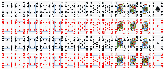
Free Images: "bestof:Notable mutations.png Příklady klinicky významných mutací Selection of notable mutations ordered in a standard table of the genetic code of amino acids As"
Notable mutations.svg
Notable mutations-es.svg
Codicegenetico.svg
Genetic code.svg
Aminoacids table.svg
Genetic code mk.svg
GeneticCode21-version-2.svg
Notable mutations.png
Cubic.jpg
Dimethylglycine 3D ball.png
Dimethylglycine zwitterion 3D ball.png
3-Aminoisobutyric-acid-3D-balls.png
3-Aminoisobutyric-acid-3D-spacefill.png
L-Butyrine-3D-balls.png
L-Butyrine-3D-spacefill.png
L-Butyrine-zwitterion-3D-balls.png
L-Butyrine-zwitterion-3D-spacefill.png
L-Thyronine 3D ball.png
L-Thyronine 3D spacefill.png
L-Thyronine zwitterion 3D ball.png
L-Thyronine zwitterion 3D spacefill.png
Dimethylglycine 3D spacefill.png
Dimethylglycine zwitterion 3D spacefill.png
John_Greenwood_-_Sea_Captains_Carousing_in_Surinam.jpg
L-Citrulline-3D-balls.png
L-Citrulline-3D-spacefill.png
L-Citrulline-zwitterion-3D-balls.png
L-Citrulline-zwitterion-3D-spacefill.png
L-Mimosine molecule ball.png
L-Mimosine molecule spacefill.png
L-Mimosine zwitterion ball.png
L-Mimosine zwitterion spacefill.png
Everything_for_the_garden_(16365135166).jpg
Terms of Use   Search of the Day