Click Here for More Images from iStock- 15% off with coupon 15FREEIMAGES 
Molecular model of Thymine, 1 of 4 nucleobases to form DNA
The Raf/MEK/ERK (MAPK) propagates growth signals in the cell. Misregulation can lead to cancer.\nThe red dots on the active EGFR are phosphorylated tyrosines. PDB entries: 1egf, 1nql, 1m17, 2jwa, 3njp, 2gs6, 1gri, 1xd2, 3ksy, 5p21, 6xi7, 6q0j, 2y4i,
3D cartoon and Gaussian surface models, chain id color scheme, PDB 2y73, white background
Top view of various kinds of vegan protein sources like tofu, tempeh, soy beans, soy milk, mushrooms, wakame, lentils, peanuts, spinach and chick peas. All the objects are at the borders of the image and at the center is written \
Different fresh products and paper with Tyrosine chemical formula on white wooden table, flat lay. Sources of essential amino acids
Photomicrograph of filamentous blue-green algae, Oscillatoria species. Each band along the stand is one cell. This filament is very short probably having recently parted from a longer one. Filaments are typically many times longer. This algae is mobile, it moves and twists through the water. San Francisco Bay, California, USA. Live specimen. Wet mount, 40X objective, transmitted brightfield illumination. Note - motion blur of live specimen, very shallow depth of field, chromatic aberration and uneven focus are inherent in light microscopy.
DNA Helix - 3d rendered image. Genetic research concept. Complex spiral structure illustration. Medical, science, genetic biotechnology, gene cell, chromosome idea. DNA strands on dark background . Science nanotechnology. Hologram view. HUD, FUI, SEM
DNA mutation, a mutation is a change in a DNA sequence, mutations can result from DNA copying mistakes made during cell division.
Food containing natural iron. Fe: Liver, avocado, broccoli, spinach, parsley, beans, nuts, on a white background. Top view.
anaplastic thyroid carcinoma with heterologous differentiation (leiomyosarcoma)
3D cartoon and Gaussian surface models, PDB 5fwk, white background
Abstract Genetics Disease - 3d rendered image.
A GTP-bound G-alpha protein (blue) binds to and activates the enzyme adenylyl cyclase, which produces then cyclic AMP spreading a signal through the cell.
Liver Fluke Section under light microscope with white background
DNA Strings On White Background. Medical Research, Gene Engineering And Biotechnology Concept
In vitro bacterial culture
CRISPR-Cas9\nproteins recognize and cut foreign pathogenic DNA
Myxoid liposarcoma.  Myxoid/round cell liposarcoma, or MRCLS, is one of several types of liposarcoma. Liposarcoma is a rare cancer that grows in the cells that store fat in the body. MRCLS usually grows in the arms and legs. These tumors grow slowly, and they can spread to other parts of the body
Products containing Vitamin B3, Niacin. 3D rendering isolated on white background
Inside protein molecule
Immunotherapy of Alzheimer's disease: Monoclonal antibody (light blue) bound to an amyloid beta peptide (orange),  that is aggregated to amyloid beta fibrils, which build up amyloid plaques.
Chain or ribbon diatom, a type of algae.
Anatomical model of large intestine and different organic products on white wooden background, flat lay. Space for text
Mobile Network Data Technology, Global Communication, Speed
Histological Cerebellum human, Sympathetic ganglion human And Spinal cord human under the microscope for education.
Wilms tumor (also called Wilms' tumor or nephroblastoma) is a type of childhood cancer that starts in the kidneys. It is the most common type of kidney cancer in children.
3D cartoon and Gaussian surface models, PDB 4bci, white background
29 October Republic Day of Türkiye text on a red shiny sphere with a partial Turkish flag on white background. Easy to crop for all your print sizes and social media needs.
Vintage photo, Ingredients or products containing vitamin B1 and dietary fiber, natural sources of minerals, healthy lifestyle and nutrition
Close-up shot of a petri dish for checking clear zone an antibiotic in the laboratory.
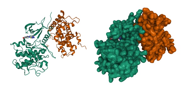
Free Images: "bestof:PDB 1nh3 EBI.jpg Multi-domain proteins alpha and beta Eukaryotic DNA topoisomerase I N-terminal DNA-binding fragment Eukaryotic DNA topoisomerase I N-terminal"
Camptothecin binding.svg
Gene2-plain-norsk.svg
Animal mitochondrion diagram it.svg
Animal mitochondrion diagram en (edit).svg
Endomembrane system diagram en (edit).svg
Animal mitochondrion diagram en.svg
Animal mitochondrion diagram bn.svg
PDB 1nh3 EBI.jpg
PDB 1k4t EBI.jpg
PDB 1rrj EBI.jpg
PDB 1rr8 EBI.jpg
PDB 1a36 EBI.jpg
PDB 1seu EBI.jpg
PDB 1t8i EBI.jpg
PDB 1k4s EBI.jpg
PDB 1lpq EBI.jpg
PDB 1r49 EBI.jpg
PDB 1ois EBI.jpg
PDB 1a31 EBI.jpg
PDB 1a35 EBI.jpg
PDB 1ej9 EBI.jpg
PDB 1sc7 EBI.jpg
PDB 1tl8 EBI.jpg
PDB 1bjt EBI.jpg
PDB 1bgw EBI.jpg
PDB 2rgr EBI.png
PDB 1vcc EBI.jpg
PDB 1mw9 EBI.jpg
PDB 1cy9 EBI.jpg
PDB 1mw8 EBI.jpg
PDB 1ecl EBI.jpg
PDB 1cyy EBI.jpg
PDB 1cy2 EBI.jpg
PDB 1cy1 EBI.jpg
PDB 1cy7 EBI.jpg
PDB 1cy8 EBI.jpg
PDB 1cy0 EBI.jpg
PDB 1cy4 EBI.jpg
PDB 1cy6 EBI.jpg
PDB 1kl9 EBI.jpg
PDB 1q8k EBI.jpg
PDB 1q46 EBI.jpg
PDB 2aho EBI.jpg
PDB 2ba0 EBI.png
PDB 2je6 EBI.png
PDB 2jea EBI.png
PDB 2nn6 EBI.png
PDB 1a41 EBI.jpg
PDB 1yua EBI.jpg
PDB 1rz4 EBI.jpg
PDB 2iwx EBI.jpg
PDB 2brc EBI.jpg
PDB 1zwh EBI.jpg
PDB 1amw EBI.jpg
PDB 1zw9 EBI.jpg
PDB 1ah6 EBI.jpg
PDB 2fxs EBI.jpg
PDB 1am1 EBI.jpg
PDB 2bre EBI.jpg
PDB 1ah8 EBI.jpg
PDB 1bgq EBI.jpg
PDB 1a4h EBI.jpg
PDB 2iws EBI.jpg
PDB 2iwu EBI.jpg
PDB 1uyl EBI.jpg
PDB 1byq EBI.jpg
PDB 2byi EBI.jpg
PDB 1yer EBI.jpg
PDB 1yc1 EBI.jpg
PDB 1osf EBI.jpg
PDB 1yc4 EBI.jpg
PDB 2byh EBI.jpg
PDB 1uy6 EBI.jpg
PDB 2h55 EBI.jpg
PDB 1uy7 EBI.jpg
PDB 1yet EBI.jpg
PDB 2cdd EBI.jpg
PDB 2uwd EBI.jpg
PDB 1uyi EBI.jpg
PDB 1uye EBI.jpg
PDB 1uyd EBI.jpg
PDB 1uy9 EBI.jpg
PDB 1uy8 EBI.jpg
PDB 2ccs EBI.jpg
PDB 1uyg EBI.jpg
PDB 2fwy EBI.jpg
PDB 2bz5 EBI.jpg
PDB 1uyc EBI.jpg
PDB 1yc3 EBI.jpg
PDB 2bsm EBI.jpg
PDB 2fwz EBI.jpg
PDB 1uyk EBI.jpg
PDB 1uyf EBI.jpg
PDB 1yes EBI.jpg
PDB 2cct EBI.jpg
PDB 1uyh EBI.jpg
PDB 1uym EBI.jpg
PDB 2ccu EBI.jpg
PDB 2gqp EBI.jpg
PDB 1qy5 EBI.jpg
Terms of Use   Search of the Day