Click Here for More Images from iStock- 15% off with coupon 15FREEIMAGES 
Old illustration of a typical cell from a 100+ year old dictionary showing all the different parts of a cell
forming of underskin cellulite illustration
Osteoporosis is a systemic skeletal disorder characterized by increased fragility and deterioration of the bone tissue. Osteoporosis is caused by an imbalance of osteoblast and osteoclast activity.
Blastomycosis is a fungal infection caused by inhaling Blastomyces dermatitidis spores. If it involves only the lungs, it is called pulmonary blastomycosis. Only about half of people with the disease have symptoms, which can include fever, cough, night sweats, muscle pains, weight loss, chest pain, and feeling tired.
superior view of heart showing opening of aorta, superior vena cava, pulmonary trunk with related valve
Photomicrograph of midge fly, Chironomidae family, eggs hatching. Eggs are encased in a gelatinous material that attracts debris. Live specimen. Larva about 0.5 mm long. Wet mount, 10X objective, transmitted brightfield illumination.
plant Zea Stem C.S. under light mircoscope with white background
Breast tissue adenocarcinoma immunohistochemistry. Estrogen receptors.
Paps smear analysis: Superficial squamous cell, metaplastic squamous cell, koilocytes cell. HPV related change
Chemical formula of Progesterone
Old anatomical model of spinal column, vertebral discs and bodies and spinal processes
Epithelioid mesothelioma is a cancer subtype caused by asbestos. It is the most common mesothelioma cell type, accounting for 50% to 70% of cases.
How a lithium ion battery works, 3d render, section. Battery charging and discharging. Ions flow from the anode to the cathode separated by a liquid electrolyte as the battery discharges energy
abstract imagery of a fusion of colors and surfaces
3D cartoon and Gaussian surface models, PDB 6gu2, white background
plant rape flower cross section under light mircoscope with white background
After dopamine binding, the dopamine receptor changes his shape and binds the inactive G protein.The G protein expels its GDP molecule replacing it by GTP. and falls into two pieces. The Galpha subunit binds to and activates the enzyme adenylyl cyclase, which produces then cyclic AMP spreading a signal through the cell.
Chemical formula of Aldosterone
Hand Drawn of Human Anatomy Cranial Bones on Chalkboard
Photomicrograph of flatworm,
the image showing fossa ovalis, crista terminalis, musculi pectinati, heart valves and inter-arterial septum
Squash preparation of stained onion root tip cells showing various stages of mitosis. Differential interference contrast optics on a Leica microscope, Image taken with the X40 objective lens.  Depth of focus is extremely shallow (just a few microns).  Differential interference contrast results in an optical slice horizontally through the specimen providing maximum possible resolution at this magnification and good contrast. Chromatic abberation is inevitable in light microscopy, but has been kept to a minimum in this image.
3D cartoon and Gaussian surface models, chain id color scheme, PDB 2y73, white background
Mesenchymal dysplasia. Site: Placenta
3D Illustration Concept of Vertebral Column of Human Skeleton System Anatomy
Chemical formula of Testosterone
Leiden leaf c.s. under microscope
The lumbar vertebrae are, in human anatomy, the five vertebrae between the rib cage and the pelvis .
Abstract technological design of an artificial molecular structure in many colors. Horizontal wide composition.
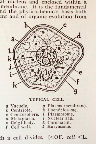
Free Images: "bestof:Plakohypaphorine F.svg diagram of the alkaloid Plakohypaphorine F own 2007-05-03 Eloil Indole alkaloids"
Paeonia suffruticosa white070503.jpg
Paeonia suffruticosa red070503B.jpg
Timeline energy companies NL.png
Plakohypaphorine A.svg
Plakohypaphorine B.svg
Plakohypaphorine C.svg
Plakohypaphorine D.svg
Plakohypaphorine E.svg
Plakohypaphorine F.svg
Brassinin (small).svg
Lespedamine.svg
Bulbocapnine.svg
5-Bromo-DMT.svg
2-Hydroxy-NMT.svg
Canthinone.svg
Convolutindole A.svg
Monoterpenoid indole alkaloids 1 ru.svg
Dauricine.svg
Fumitremorgin C.svg
Tryprostatin A.svg
Boldine.svg
Jatrorrhizine.svg
Brucine.svg
Toxiferin.svg
Harmalin.svg
Ajmaline structure.svg
Tropane family ru.svg
(+) Stephacidin A.svg
Ajmalicin.svg
Mitragynine.svg
Reserpsäuremethylester.svg
Methyl ketoreserpate.svg
Ajmalicine.svg
Brevianamide A.svg
Brevianamide B.svg
Heyneanine.svg
Apparicine.svg
Pergolide.svg
18-Methoxycoronaridine.svg
Yuehchukene.svg
Birnbaumin A.svg
Birnbaumin B.svg
Vindoline.svg
Deacetylvindoline.svg
Desacetoxyvindoline.svg
16-Hydroxytabersonine.svg
Tabersonine.svg
4,21-Dehydrogeissoschizine.svg
Preakuammicine.svg
Stemmadenine.svg
Dehydrosecodine.svg
Camalexin.svg
Aspidospermidine.svg
Pergolid Structural Formulae V.1.svg
Fellutanines A,B,C,D.svg
Amaranthin.svg
N-Methyltryptamine.svg
Yohimban - Yohimban.svg
Reserpinine.svg
Conodurine.svg
16-methoxytabersonine.svg
Fellutanine A and B.svg
Fellutanine A-D.svg
Yohimban num.svg
Norbaeocystin.svg
Rauwolscine.svg
3-Hydroxy-16-methoxy-2,3-dihydrotabersonine.svg
Rescinnamine.svg
Coronaridine.svg
(+)-K252a.svg
(1S)-1-methyl-2,3,4,9-tetrahydro-1H-pyrido-3,4-b-indole.svg
Voacangine.svg
Ibogamine.svg
Cisatracurium.svg
Ergometrine.svg
Doxacurium chloride.svg
Doxacurium.svg
Aspidophytine.svg
Ethocybin.svg
Bufotenine.svg
Tryptophan decarboxylation.svg
Villalstonine.svg
Tetrahydroharmine structure.svg
Diagram Needed.svg
Indol2.svg
Tryptophan decarboxylation (en).svg
FischerIndoleSynthesisMechanism.svg
Indole-3-propionamide.svg
Indol (vzorec).svg
Indolepropionamide.svg
Indol.svg
LMFMultilingualNotationsfleuve.svg
LMFMultilingualNotationsdevelop.svg
Indole numbered.svg
Indolylglycine.svg
Damascenin.svg
AND Gate diagram.svg
Li2CO3.svg
Bukkake.svg
Himbacine.svg
Terms of Use   Search of the Day