Click Here for More Images from iStock- 15% off with coupon 15FREEIMAGES 
Water algae cells - microscope magnification
Close up of cristal beads
The Microscopic World. Onion epidermis with cells.
Microscopic image of Microscopic image of nymphaea of aqustio stem
Host cells with spores (mold) are inside wood under the microscope for education.
Round, green chloroplasts in plant cells of anacharis or waterweed, Egeria densa. Chloroplasts carry chlorophyll which makes them green. These chlorplasts actually circulate around within each cell. Live specimen. Wet mount, 40X objective, transmitted brightfield illumination. Note - very shallow depth of field, chromatic aberration and uneven focus are inherent in light microscopy.
linden stem (Tilia platyphyllos) cross section under the microscope showing phloem, vascular cambium, medullary ray and pith - optical microscope x32 magnification
corn stem micrograph with dye
view of vegetal cells through a 1000x microscope lens
plant Tilia Stem C.S. under light mircoscope with white background
Lime trunk
Industrial plant, paper mill, with smoking chimneys viewed directly from above.
Cross-section Dicot, Monocot and Root of Plant Stem under the microscope for classroom education.
Close-up shot of a petri dish for checking an antibiotic in the laboratory.
Leiden leaf c.s. under microscope
Aerial photo of industrial site.
Pine mature wood cross section. Light microscope slide with microsection of an evergreen conifer in the genus Pinus. Plant anatomy. Biology. Photo.
plant rape flower cross section under light mircoscope with white background
A giant power station in the United Kingdom, seen from the air.
Petrochemical plant with many large storage tanks, pipelines and chimneys, surrounded with green forest, viewed from above.
The study of plant tissues under the microscope in the laboratory.
Microscopic image of Microscopic image of nymphaea of aqustio stem
Industrial oil refinery gas flare, safety device for burning off unwanted flammable gasses, Immingham
Close-up shot of a petri dish for checking an antibiotic in the laboratory.
A closeup shot of a laboratory worker examining a green substance on a petri dish while conducting coronavirus research
Scientist looking at a fungal culture at the laboratory - medical diagnostic concepts
Plant tissue  under a microscope
Abstract particles
Cross-section leaf Plant of under the microscope for classroom education.
A large oil refinery along the shores of the Mississippi River in Louisiana near the Gulf of Mexico.
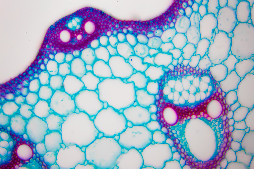
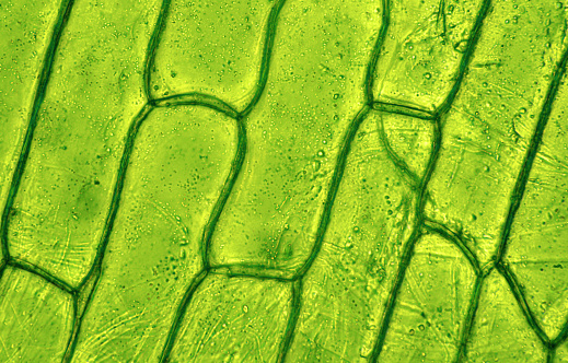
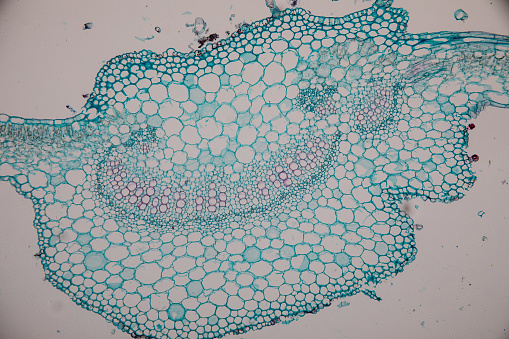
Free Images: "bestof:Plant cell structure svg uk.svg uk Ст� укту� а � ослинної клітини 2011-06-26 Image Plant cell structure svg svg LadyofHats Mariana Ruiz"
Plant cell structure svg uk.svg
Animal cell structure uk.svg
Plant cell structure svg-zh-cn.svg
Plant cell structure svg-de.svg
Plant cell structure svg pl.svg
Plant cell structure tr.svg
Plant cell structure svg hu.svg
Plant cell structure svg-id.svg
Plant cell structure svg 2.svg
Plant cell structure svg zh-hant.svg
Plant cell structure svg labels.svg
Struttura della cellula vegetale.svg
Plastids types uk.svg
Turgor pressure on plant cells diagram-uk.svg
Cellular tight junction keys.svg
Plant cell structure fr.svg
Plant cell structure pt.svg
Plant cell wall diagram-uk.svg
Adherens Junctions structural proteins-fr.svg
Adherens Junctions structural proteins.svg
Diagram human cell nucleus uk.svg
Average prokaryote cell- uk.svg
Desmosome cell junction uk.svg
Endomembrane system diagram en (edit).svg
Animal mitochondrion diagram en.svg
Animal mitochondrion diagram cs.svg
Animal mitochondrion diagram bn.svg
Animal mitochondrion diagram en (edit).svg
Plant cell structure-en.svg
Plant cell structure no text-2.svg
Animal cell structure pl.svg
Complete neuron cell diagram uk.svg
Animal cell structure numbered version.svg
Animal cell structure sv.svg
Animal cell structure id.svg
Desmosome cell junction it.svg
Desmosome cell junction en.svg
Plastids types en.svg
Bacterial morphology diagram-es.svg
Animal cell structure no text.svg
Cell junction simplified en.svg
Bacterial morphology diagram.svg
Animal cell structure en.svg
Animal cell structure de.svg
Average prokaryote cell- en.svg
Bacterial morphology diagram fr.svg
Plastids types-fr.svg
Plastids types it.svg
Plant cell structure svg vacuole.svg
Plant cell structure cs.svg
Gap cell junction-uk.svg
Cell junction simplified uk.svg
Plant cell structure svg 2 sl.svg
Plant cell structure svg-es.svg
Plant cell structure-gl.svg
Animal cell structure pt.svg
Connexon and connexin structure gl.svg
Cellular tight junction-uk.svg
Angiosperm life cycle diagram uk.svg
Adherens Junctions structural proteins-ja.svg
Plastids types cs.svg
Culex mosquito life cycle uk.svg
Culex pipiens diagram uk.svg
Plastids types es.svg
Connexon and connexin structure fr.svg
Connexon and connexin structure.svg
Chloroplast diagram (zh-cn).svg
Thyroid hormone synthesis-it.svg
Chloroplast diagram.svg
Thyroid hormone synthesis.svg
Plant cell wall diagram mk.svg
NK-cell activation.svg
Komorkaroslinna.svg
Cell membrane detailed diagram ua.svg
Average prokaryote cell numbered.svg
Average prokaryote cell- fr.svg
Schema pflanzliche Zellwand.svg
Average prokaryote cell-pl.svg
Diagram human cell nucleus oc.svg
Cell membrane detailed diagram pt.svg
Prokaryote cell diagram he.svg
Diagram human cell nucleus (zh-cn).svg
Diagram human cell nucleus tr.svg
Average prokaryote cell- da.svg
Gap cell junction keys.svg
Average prokaryote cell- ru.svg
Cell junction simplified zh traditional.svg
Gap cell junction-cs.svg
Diagram human cell nucleus cs.svg
Diagram human cell nucleus pt.svg
Gap cell junction-de.svg
Diagram human cell nucleus sl.svg
Gap cell junction it.svg
Prokaryote cell diagram cs.svg
Endomembrane system diagram cs.svg
Diagram human cell nucleus hu.svg
Gap cell junction-es.svg
Cell junction simplified it.svg
Average prokaryote cell- id.svg
Fagocytoza.svg
Terms of Use   Search of the Day