Click Here for More Images from iStock- 15% off with coupon 15FREEIMAGES 
Y-Chromosomes with DNA carrying the genetic code. Genetics concept, medicine concept. Future, genetic mutations. Changing the genetic code at the biological level, 3D illustration
anaplastic thyroid carcinoma with heterologous differentiation (leiomyosarcoma)
Dna structure on science background. 3d illustration
background
Microalgae under microscopic view, green algae, cyanobacteria, phytoplankton, diatom, algae mix collage background
Squash preparation of  onion root tip cells stained to reveal chromosomes,  Bright field illumination, light microscopy.  This image was taken with the X40 objective lens.  Depth of focus is extremely low at this magnification.  Smaller apertures give greater depth of focus, but image quality deteriorates and resolution is greatly reduced.  The aperture in this image was optimised for contrast and resolution. Chromatic abberation is inevitable in light microscopy, but has been kept to a minimum in this image.
Thyroid cancer: Microscopic image of Metastatic papillary carcinoma of thyroid. Reactive change. Lymph node carcinoma.
Abs Virus - 3d rendered image like colorized scanning electron micrograph. Scene showing virus (yellow)  emerging from infected cell.\nConcept of coronavirus that’s causing a global pandemic. Covid-19, Sars-cov-2 alpha, betta, gamma, delta, lambda variant, 2019-nCoV, Coronavirus.\nSEM, TEM, hologram view.
High Vaginal Swab (HVS) Gram Stain: Photomicrograph showing Bacterial vaginosis (BV).
Wilms tumor (also called Wilms' tumor or nephroblastoma) is a type of childhood cancer that starts in the kidneys. It is the most common type of kidney cancer in children.
Illustration showing chromosomes with highlighted telomeres
Liver Fluke Section under light microscope with white background
Nodularia sp. toxic algae under microscopic view, nitrogen-fixing cyanobacteria, cyanotoxin, harmful to humans, x40
Abstract Genetics Disease - 3d rendered image.
Genetics background. 3D render.
Immunotherapy of Alzheimer's disease: Monoclonal antibody (light blue) bound to an amyloid beta peptide (orange),  that is aggregated to amyloid beta fibrils, which build up amyloid plaques.
Photomicrograph of filamentous blue-green algae, Oscillatoria species. Each band along the stand is one cell. This filament is very short probably having recently parted from a longer one. Filaments are typically many times longer. This algae is mobile, it moves and twists through the water. San Francisco Bay, California, USA. Live specimen. Wet mount, 40X objective, transmitted brightfield illumination. Note - motion blur of live specimen, very shallow depth of field, chromatic aberration and uneven focus are inherent in light microscopy.
Backgrounds of Bacteria and Mold under the microscope in Lab.
Myxoid liposarcoma.  Myxoid/round cell liposarcoma, or MRCLS, is one of several types of liposarcoma. Liposarcoma is a rare cancer that grows in the cells that store fat in the body. MRCLS usually grows in the arms and legs. These tumors grow slowly, and they can spread to other parts of the body
Light microscope micrograph showing mitosis in onion cells of the root meristem. At bottom, is a cell in anaphase. On the top right corner, a prophase.
PAPS Smear study of a young women under microscopy showing Atrophic changes in uterus which is unusual at young age.
Squash preparation of  onion root tip cells stained to reveal chromosomes,  Bright field illumination, light microscopy.  This image was taken with the X40 objective lens.  Depth of focus is extremely low at this magnification.  Smaller apertures give greater depth of focus, but image quality deteriorates and resolution is greatly reduced.  The aperture in this image was optimised for contrast and resolution. Chromatic abberation is inevitable in light microscopy, but has been kept to a minimum in this image.
Isolated Poly Protein G with Detection Antibodies to enhance Immunoassays sensitivity 3d rendering
Genetic research abstract - Colorectal cancer
A new antibiotic (zosurabalpin, red) traps lipopolysaccharide (orange) in the intermembrane transporter of the Gram-negative bacterium Acinetobacter baylyi
plant Leaf of Nerium indicum T.S. under light mircoscope with white background
Algaes under microscopic view
Epithelioid mesothelioma is a cancer subtype caused by asbestos. It is the most common mesothelioma cell type, accounting for 50% to 70% of cases.
Lily anther c.s. under microscope
Close-up shot of a petri dish for checking an antibiotic in the laboratory.
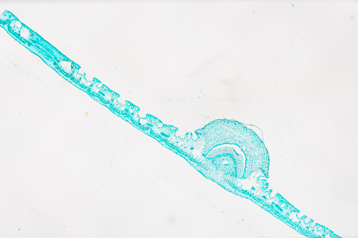
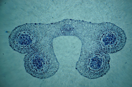
Free Images: "bestof:Polyten chromosome.jpg Polyten chromosome from fly salivary gland Own 12/01/06 LPLT User LPLT/Credits Polytene chromosomes Drosophila"
Chromosom2.svg
Chromosome mutations-ar.svg
Chromosomes mutations-en.svg
Chromosomes mutations-mk.svg
Digestive system diagram en.svg
Chromosome fr.svg
Chromosome zh.svg
Chromosome zh-tw.svg
Chromosome Conformation Capture Technology (zh-cn).svg
HaplogroupsCaucasus.svg
Chromosom.svg
Hox cluster.svg
HLA.svg
Autodominant.svg
Speciation experiment.svg
Drosophila speciation experiment.svg
Drosophila speciation experiment NL.svg
Polyten chromosome.jpg
Drosophila-chromosome-diagram.jpg
Human chromosomesXXY02.png
Chromosome Conformation Capture Technology.jpg
Human chromosomesXXY01.png
Drosophila polytene chromosomes.jpg
Chromosome_12.jpeg
Map of Chromosome 12.png
Chromosome_20.jpeg
PPHSimage01.jpg
Linkage.png
87AC polytene heatshock puffs-1-.jpg
Basilica San Vital di Ravenna.JPG
Lycée Montaigne 01.jpg
Over-represented Ligation Junctions.jpg
Patio grande mosquee de paris.jpg
Plaque des Arenes de Lutece.jpg
Trieste central station.JPG
Sorbonne, rue Victor-Cousin.jpg
Maison de Marin-Marie a Chausey.jpg
Fontaine_Sainte-Barbe_du_Faouët_par_Henri_Barnoin.JPG
Vieille_Bretonne_(1910)_-_Henri-Jules_Guinier.JPG
City Life Theme Steve Reich - 2.png
Genelocation2.png
Breton_(1910)_Charles_Rivière.JPG
Chromosome_21.png
Mappa Linea B - Metropolitana di Roma.PNG
Human male karyotpe high resolution - XY chromosome cropped.JPG
Flemming1882Tafel1Fig14.jpg
Hemophilia_01.gif
Hemophilia_02.gif
Chromosoom Dutch text.png
Genes and bases on chromosomes.png
Drosophila-melanogaster-Nauener-Stadtwald-03-VII-2007-12.jpg
Chromosome Spanish text.png
Meiosis II Non Disjunction.jpg
Pre-RC.png
E1b1bRoute.png
La Danse de Carpeaux.jpg
Chromosome Turkish Text.png
Chromosom.png
Loss of Heterozygosity.png
PCA of Haplogroup J from 6 STRs.png
Loss of heterozygosity.PNG
PCA of Haplogroup J using 37 STRs.png
Log2R.jpg
Carettochelys insculpta LA zoo.jpg
Guadeloupe_sur_planisphère_de_Cantino_1502.jpg
Speciation experiment.png
TMRCAs-compared.PNG
Terms of Use   Search of the Day