Click Here for More Images from iStock- 15% off with coupon 15FREEIMAGES 
Bacterial multidrug efflux pumps (E-CmeB) expel antibiotics  from inside the bacterium (bottom): ciprofloxacin (yellow), chloramphenicol (violet),  ampicillin (red).
Microscopic image of Cucurbita
3D cartoon and Gaussian surface models. PDB 3kqg, chain id color scheme, white background.
Human embryonic stem cells
Acetylcholine receptors in the lipid layer of cell membrane.
Phospholipid bilayer membrane structures that make up living cells and their contents. This is an image of membrane proteins in the membrane.\nThis image, which looks like a plastic model, evokes biology and biochemistry. It can be used as a cover for magazines, textbooks and presentation materials.\nThis image was not created by AI.
Cell Membrane and Molecules, 3d rendering. Computer digital drawing.
ATP Synthase Protein - 3d rendered image. Medical microbiology concept.  Structure and conformational states of the bovine mitochondrial ATP (Adenosine triphosphate) synthase. Chemical energy is synthesized in eukaryotic cells primarily. Hydrolase. Hologram view. HUD, FUI
The calcitonin gene related peptide (yellow) binds to its receptor (blue) on neurons and smooth muscle cells of cerebral blood vessels, activating a signal cascade through G-proteins (dark blue) in this cells that  leads to a dilatation of the blood vessels (vasodilatation).
Cells in the body examined by endoscope
SARS-CoV-2 ORF3a is a putative viral ion channel implicated in autophagy inhibition, inflammasome activation and apoptosis. 3D cartoon and Gaussian surface models, based on PDB 7kjr, white background.
plant Pine Stem C.S. under light mircoscope with white background
Cross-section illustration of human skin, composed of three primary layers: epidermis, dermis and subcutis.
3d rendering of Easter eggs background
Cell Membrane and Molecules, 3d rendering. Computer digital drawing.
Covid-19 Coronavirus Variants Omicron Delta Gamma Beta Alpha Pandemic Virus Disease Mutation
After dopamine binding, the dopamine receptor changes his shape and binds the inactive G protein.The G protein expels its GDP molecule replacing it by GTP. and falls into two pieces. The Galpha subunit binds to and activates the enzyme adenylyl cyclase, which produces then cyclic AMP spreading a signal through the cell.
Endosomes - 3d rendered image of Endosomes are membrane-bound organelles. They are parts of endocytic membrane transport pathway.  Endomembrane system in cells. Bilayer Lipid Membrane. Abstract medical illustration. Microscopic view. Microbiologic research concept.
Proteasomes are molecular machines for breaking down proteins called proteolysis, only target proteins that have been marked for destruction called ubiquitin to the target protein, 3d rendering
A conceptual image of an abstract network of multi-coloured organic strands arranged in a non regular pattern all connected together in a complex manner. The network is made from smaller multi-coloured squares and polyhedra, in vivid colours against a pastel toned background. Shows background and foreground elements to the structure.
3D cartoon and Gaussian surface models, chain id color scheme, PDB 7t3a, white background
Digital background depicting innovative technologies in (AI) artificial systems, neural interfaces and internet machine learning technologies
Artificial Intelligence: Simplified image of the principles of convolutional neural networks with input, hidden, output layers. It classifies geometric forms - in this case a cube
Human Morula Cell, Solid Ball of Cells Resulting from Division
HER2 (light blue)  locked in the inactive conformation by Trastuzumab (left) and Pertuzumab. Trastuzumab has drastically improved the prognosis of HER2-positive breast cancer patients.
plant rape flower cross section under light mircoscope with white background
Coxsackie virus internal structure, 3d illustration set isolated on white
Endosomes - 3d rendered image of Endosomes are membrane-bound organelles. They are parts of endocytic membrane transport pathway.  Endomembrane system in cells. Bilayer Lipid Membrane. Abstract medical illustration. Microscopic view. Microbiologic research concept.
Spike glycoprotein homotrimer, a main target for neutralization antibody, to bind its receptor, ACE2, and mediate membrane fusion and virus entry. 3D cartoon and Gaussian surface models, white background.
Particles, medical concept
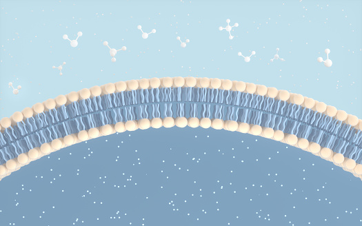
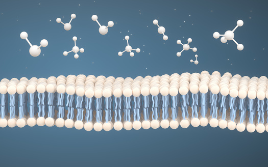
Free Images: "bestof:Scheme simple diffusion in cell membrane-de.png Scheme simple diffusion in cell membrane-de svg Einfaches Modell der Diffusion an einer Zellmembran DIe"
Scheme simple diffusion in cell membrane-de 02.svg
Scheme simple diffusion in cell membrane-de.svg
Scheme simple diffusion in cell membrane-zh (zh-cn).svg
Scheme simple diffusion in cell membrane-zh (zh-tw).svg
Scheme simple diffusion in cell membrane-en.svg
Scheme simple diffusion in cell membrane-es.svg
Scheme simple diffusion in cell membrane-fr.svg
Scheme simple diffusion in cell membrane-ca.svg
Scheme simple diffusion in cell membrane-pl.svg
Scheme facilitated diffusion in cell membrane-zh.svg
Scheme facilitated diffusion in cell membrane-zh (zh-tw).svg
Scheme facilitated diffusion in cell membrane-en.svg
Scheme facilitated diffusion in cell membrane-es.svg
Scheme facilitated diffusion in cell membrane-ca.svg
Scheme facilitated diffusion in cell membrane-eo.svg
Scheme facilitated diffusion in cell membrane-fr.svg
Scheme secundary active transport-en.svg
Scheme secundary active transport-heb.svg
Scheme secundary active transport-pl.svg
Scheme sodium-potassium pump-de.svg
Scheme sodium-potassium pump-cs.svg
HEPA Filter diagram de.svg
Complement pathway.svg
Deposition scheme german.svg
Diocèse de Saint-Dié.svg
Gray5_de.svg
Gray5_nl.svg
Doric_Chiton.svg
Medial axis example 2d.svg
Phylogenetic_tree-de.svg
ShockleyQueisserFullCurve (DE).svg
ShockleyQueisserBreakdown2 (DE).svg
Diocèses (France).svg
Diffusion (schematic).svg
cell_simple.svg
phone-cell-phone-simple-outline-312200.svg
Entropy and diffusion (de).svg
Simple heat emitting circuit.svg
Euglenid pellicula scheme.svg
Diffusionsexperiment.svg
1829 Braille cell numbering.svg
Hashlink timestamping.svg
head-water-chemical-diagram-simple-38071.svg
cellphone-mobile-phone-155337.svg
Simple aldol reaction.svg
card-cell-icons-mobile-phone-sim-1294054.svg
camera-cell-flash-icons-mobile-1294053.svg
Simple DirectMedia Library de.svg
List icon.svg
Cell membrane detailed diagram de.svg
keyhole-keyhole-bed-permaculture-1609542.svg
Schema pflanzliche Zellwand.svg
Desmosome scheme.svg
Scheme human hand bones-en.svg
Complete neuron cell diagram de.svg
Capacitor resistor series.svg
Scheme human hand bones-numbers.svg
Scheme human hand bones-de.svg
ExampleOfBulkPhotovoltaicEffect.svg
Major events in mitosis-gl.svg
Gray's_7_(ovum_maturation).svg
Plant cell structure svg 2.svg
Scheme sodium-potassium pump-ja.svg
Plant cell structure-en.svg
Plant cell structure svg labels.svg
HIV-timecourse simple.svg
SiteLaboAndra1988.svg
SiteLaboAndra1993.svg
Province ecclésiastique de Tours.svg
Archidiocèse de Tours.svg
Archidiocèse de Rennes.svg
Archidiocèse de Dijon.svg
Province ecclésiastique de Dijon.svg
Diocèse du Mans.svg
Archidiocèse de Lyon.svg
Province ecclésiastique de Lyon.svg
Archidiocèse de Marseille.svg
Province ecclésiastique de Marseille.svg
Archidiocèse de Lille.svg
Province ecclésiastique de Lille.svg
Diocèse d'Autun.svg
Archidiocèse de Reims.svg
Province ecclésiastique de Reims.svg
Diocèse de Beauvais.svg
Archidiocèse de Clermont.svg
Province ecclésiastique de Clermont.svg
Diocèse de Nantes.svg
Archidiocèse de Besançon.svg
Archidiocèse de Bordeaux.svg
Province ecclésiastique de Bordeaux.svg
Archidiocèse de Toulouse.svg
Province ecclésiastique de Toulouse.svg
Archidiocèse de Strasbourg.svg
Diocèse de Metz.svg
Archidiocèse de Rouen.svg
Province ecclésiastique de Rouen.svg
Diocèse de Nice.svg
Province ecclésiastique de Montpellier.svg
Archidiocèse de Montpellier.svg
Province ecclésiastique de Poitiers.svg
Terms of Use   Search of the Day