Click Here for More Images from iStock- 15% off with coupon 15FREEIMAGES 
A ball and stick model of Human Insulin. Isolated on black.
A plasmid is a small circular DNA molecule found in bacteria and some other microscopic organisms 3d rendering
Dna structure on science background. 3d illustration
DNA Molecule: a 3D illustration of the double helix structure reflecting the genetic code. This 3D illustration is a visualization of the DNA molecule showing its characteristic double helix structure. The model reflects the major components of DNA such as the nitrogenous bases (adenine, thymine, guanine, and cytosine), sugars, and phosphate groups that together form the genetic code. This illustration can be used for educational purposes, scientific publications or medical advertising to demonstrate the complexity and beauty of the molecule responsible for heredity and the development of life on Earth.
ribonicleic acid chain from which the deoxyribonucleic acid or DNA is composed - 3d illustration
3d rendering of transparent molecule shape on green color background.
Bacterial multidrug efflux pumps (E-CmeB) expel antibiotics  from inside the bacterium (bottom): ciprofloxacin (yellow), chloramphenicol (violet),  ampicillin (red).
Dna structure digital concept
Abstract DNA spiral model (done in 3d)
DNA, Adenine, Cytosine, Guanine, Laboratory
Prostate Cancer: Microscopic image of prostatic tissue, adenocarcinoma, malignant neoplasm, atypical epithelial cells.
Molecular structure formula. Concept. 3D Render
Multicolored antibodies or immunoglobulin protein structures - 3d illustration
Quantum Gravity 3d rendered image of a comprehensive exploration of Quantum Gravity theories. String theory, loop quantum gravity (LOG), causal dynamical triangulation, twistor theory, gravitational waves, spacetime, gravity waves, quantum mechanics, quantum entanglement concepts.
Digital models of DNA structure with problem element on abstract blue background. 3d render
Dna structure digital concept
DNA molecules chains floating in water. Abstract dna from glass material. One Dna chain is in focus and other are not. Close up, selective focus. 3D rendering illustration
Futuristic artificial DNA strand among organic models. Medical innovation and technology concept.
Medical capsule with DNA inside, 3d rendering. 3D illustration.
Models of a DNA strand with shallow depth of field.
3d helix chromosome or Dna structure, technology science background. 3d render illustration
Illustration of DNA.
RNA And DNA concept as a deoxyribonucleic acid or a ribonucleic acid as biological molecules as a symbol of the evolution of life and genetic sequencing.
Chain of Amino Acid. Proteins consist of one or more chains of amino acids called polypeptides
Haematococcus pluvialis green and cyst algae under microscopic view, empty space - haematocyst, active and resting cells, strong antioxidant astaxanthin
3D illustration of DNA molecule: the perfect image for scientific, medical and educational projects - this breathtaking image is a realistic 3D illustration of the DNA molecule, focusing on its double helical structure. Ideal for use in scientific articles, medical presentations, educational materials and other projects related to genetics, biology and medicine. The image is high resolution and detailed, allowing you to see every strand and base pair in the DNA structure. It is also suitable for use as an illustration in textbooks, popular science articles and other materials where a visual representation of genetic material is required.
Helical DNA gene molecules on a blurred background.
Artificial Intelligence Sustainable Responsible Technology
p53 (blue) tagged with ubiquitin (yellow) is degraded into small peptides by a proteasome (violett). p53 prevents cancer formation and acts as a guardian of the genome. Mutations in the p53 gene contribute to about half of the cases of human cancer.
Texture Pattern Design on Banknote
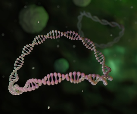
Free Images: "bestof:Telomere quadruplex.jpg en Crystal structure of parallel quadruplexes from human telomeric DNA The DNA strand circles the bases that stack together in the"
DNA chemical structure hy.svg
dna-helix-curve-science-medicine-869109.jpg
dna-helix-curve-science-medicine-869110.jpg
DNA chemical structure.svg
strand-synthesis-single-dna-human-42586.svg
Dna repair base excersion en.svg
Albero Y-DNA.svg
AsDNA chemical structure.svg
dna-biology-medicine-gene-163466.jpg
dna-helix-science-biology-research-308919.svg
DNA intercalation.svg
dna-rna-genes-blue-double-helix-297576.svg
Chromosome fr.svg
Average prokaryote cell- en.svg
Crystal facet formation.svg
Neighbor-joining Tree.svg
Neighbor-joining Tree (Editable).svg
Wu-UPGMA-Tree-Acanthobasidium-Aleurodiscus-Neoaleurodiscus.svg
Endomembrane system diagram en (edit).svg
Animal mitochondrion diagram en.svg
2ph1 pdb gallery nucleotide binding-protein-2 (MinD homolog).svg
Animal mitochondrion diagram en (edit).svg
Hepatitis B virus v2.svg
Notable mutations.svg
Bondarzewiaceae Maximum Likelihood-Tree .svg
Design_Analysis_of_the_P-38_Lightning.png
portugues-sexo_alemão-sex_ingles-sex_africano-seks_russo-секс_albanes-sex_chines-性別_bulgaro-секс_croata-spol_gales-rhyw_coreano-섹스_frances-sexe_japones-セックス_filandes-sukupuolesta_arabe-الجنس_prontos_ai_tens_em_15_linguas_a_p.jpg
Telomere quadruplex.jpg
Telomere quadruplex without fog.jpg
Ube3a.jpg
RAP1-rasmol.GIF
Crystal Structure of the Ku70 - 42.png
Crystal Structure of the Ku70 - 43.png
Crystal Structure of the Ku80 - 11.png
Crystal Structure of the Ku80 - 37.png
Crystal Structure of the Ku80 - 38.png
Crystal Structure of the Ku80 - 39.png
Crystal Structure of the Ku80 - 40.png
Crystal Structure of the Ku70 - 41.png
Crystal Structure of the Ku Heterodimer - 15.png
Crystal Structure of the Ku Heterodimer - 16.png
Crystal Structure of the Ku Heterodimer - 17.png
Crystal Structure of the Ku Heterodimer - 2.png
Crystal Structure of the Ku Heterodimer - 20.png
Crystal Structure of the Ku Heterodimer - 22.png
Crystal Structure of the Ku Heterodimer - 23.png
Crystal Structure of the Ku Heterodimer - 26.png
Crystal Structure of the Ku Heterodimer - 30.png
Crystal Structure of the Ku Heterodimer - 31.png
Crystal Structure of the Ku Heterodimer - 36.png
Crystal Structure of the Ku Heterodimer - 35.png
Crystal Structure of the Ku Heterodimer - 33.png
ACD-TPP1 rasmol.png
Quadruplex1 s.png
TRF1-DNA.png
ExonucleaseIIIfromEColi.png
Threepol.gif
Ku bound to DNA.png
Ligase rxn.png
Uracil base glycosidase.jpg
Foxp1 2KIU.jpg
Crystal Structure of the FokI endonuclease - PDB-2FOK.png
RNA pol.jpg
Crystal Structure of the FokI endonuklease - PDB-2FOK.png
1N6J.png
RecBCD crystal structure.jpg
2RAM.png
DNA_damaged_by_carcinogenic_2-aminofluorene_AF.jpg
DNA-structure-and-bases.png
Crystal Structure of Human Diamine Oxidase.png
POT1.png
SRY+DNA 1J46.png
DNA intercalation2.jpg
DNA-adduct M1G.png
Stat1 stucture.png
J-Y-DNA.jpg
Stat3 structure.png
Mitochondriaal_DNA_loa.png
DNA-internal-CpG-dinucleotide.png
PAX+ETS+DNA 1K78.png
MGMT+DNA 1T38.png
J-Y-DNA-Distribution.jpg
IL6 Crystal Structure.png
PDB 1jb7 EBI.jpg
PDB 2i0q EBI.jpg
PDB 1ph6 EBI.jpg
PDB 1ph4 EBI.jpg
PDB 1ph5 EBI.jpg
PDB 1ph8 EBI.jpg
PDB 1ph3 EBI.jpg
PDB 1pa6 EBI.jpg
PDB 1ph9 EBI.jpg
PDB 1ph1 EBI.jpg
PDB 1kix EBI.jpg
PDB 1phj EBI.jpg
PDB 1k8g EBI.jpg
PDB 1otc EBI.jpg
PDB 1ph2 EBI.jpg
Terms of Use   Search of the Day