Click Here for More Images from iStock- 15% off with coupon 15FREEIMAGES 
the tail of Aelurostrongylus abstrusus, cat lungworm under the microscope
Rare Scorpion (Arachnida) inclusion in Burmese amber, Myanmar, Cenomian, Upper Cretaceous, approximately 100 million years ago, age of dinosaurs. Image taken with extreme macro and focus stacking technique.
centropyxis aculeata (amoeboid organism) under the microscope - optical microscope x400 magnification
plant Zea Stem C.S. under light mircoscope with white background
Planarian parasite (flatworm) under microscope view.
Photomicrograph of algae, Cosmarium turpinii, a desmid. Live specimen. Wet mount, 40X objective, transmitted brightfield illumination.
Bacteria of different shapes, cocci and rod-shaped bacilli, 3D illustration in old book vintage style
Haliotis kamtschatkana, northern abalone or pinto abalone, is a species of large sea snail, a marine gastropod mollusc in the family Haliotidae, the abalones. It has been listed as \
Freshwater aquatic zooplankton and algae under microscope view
Rat tailed maggot photographed in a studio
Leiden leaf c.s. under microscope
Trematode parasite under a microscope
Planarian parasite (flatworm) under microscope view.
Liver Fluke Section under light microscope with white background
Pentatrichomonas hominis protozoan, 3D illustration. Also known as Trichomonas hominis or T. intestinalis, colonizes human large intestine, usually asymptomatic but may cause diarrhea
Close up of a House Centipede
Aquatic (fresh water) nematode living in pond water among cyanobacteria. Live specimen. Wet mount, 10X objective, transmitted brightfield illumination.
Freshwater Closterium algae (unicellular charophyte green algae) - optical microscope x200 magnification
amazing inhabitants of the microworld under a microscope
abs Lactobacillus Bulgaricus Bacteria - 3d rendered microbiology image. Medical research, health-care concept.
Eggs of helminth and Finding parasites in feces, analyze by microscope in laboratory.
Escherichia Coli (E. Coli.)
Beekeeping. Queen bee in a transport container.
Blastomycosis is a fungal infection caused by inhaling Blastomyces dermatitidis spores. If it involves only the lungs, it is called pulmonary blastomycosis. Only about half of people with the disease have symptoms, which can include fever, cough, night sweats, muscle pains, weight loss, chest pain, and feeling tired.
newly hatched freshwater fish under a light microscope, isolated on white
Natural agate surfaces, suitable for backgrounds and wallpapers.
Eggs of helminths Enterobius vermicularis containing worm larva. Threadworm which cause enterobiasis, 3D illustration
Sciaroidea insect Fossil inside Burmese Amber of Cenomanian Era, 100 million years ago, from the state of Myanmar
Tapeworm (Parasitic flatworm) of cattle and other grazing animals under the microscope for education.
plant rape flower cross section under light mircoscope with white background
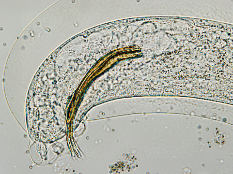
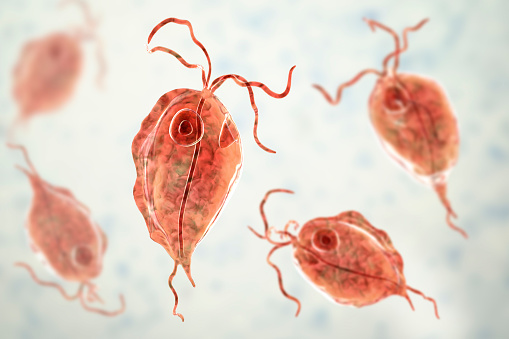
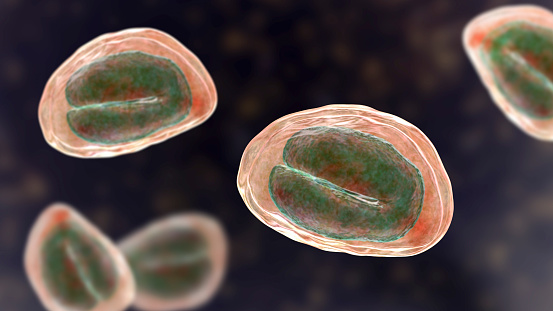
Free Images: "bestof:Trypanosoma cruzi hindgut.jpg Trypanosoma cruzi parasites in hindgut of a field-collected triatomine bugs source http //www cdc gov/ncidod/EID/vol9no1/..."
Journal_of_Emerging_Infectious_Diseases_Feb_2013.jpg
Tobacco use in the United States.svg
Toxoplasmosis life cycle en.svg
Leishmaniasis life cycle diagram en.svg
Leishmaniasis life cycle diagram fr.svg
Symptoms of ebola (vector image).svg
HIV cases by transmission category CDC 2007.svg
Hepatitis C infection by source (CDC) - en.svg
Leishmaniasis life cycle diagram ar.svg
Leishmaniasis life cycle diagram ru.svg
Toxoplasmosis life cycle de.svg
Leishmaniasis life cycle diagram-es.svg
World98.svg
TasacoraTest-World98.svg
TasacoraTest-wheelManager-World98.svg
Trypanosoma_cruzi_hindgut.jpg
Cykl_życiowy_świdrowca_amerykańskiego_CDC.JPG
Trypanosoma_cruzi_LifeCycle.gif
Trypanosoma_cruzi_LifeCycle_cs.png
Trypanosoma_cruzi_triatomid_bite1.jpg
Trypanosoma_blood.jpg
Tcruzi_IFA_positive.jpg
Trypanosoma cruzi ciclo de vida.png
Trypanosoma cruzi lifecycle dumb.png
TB8Cs2H1 snoRNA.PNG
TB10Cs4H4 snoRNA.PNG
TB11Cs4H3 snoRNA.PNG
TB3Cs2H1 snoRNA.PNG
TB9Cs4H1 snoRNA.PNG
TB11Cs5H1 snoRNA.PNG
TB8Cs4H2 snoRNA.PNG
TB10Cs2H2 snoRNA.PNG
TB6Cs1H1 snoRNA.JPG
TB6Cs1H3 snoRNA.PNG
TB10Cs1H1 snoRNA.PNG
TB9Cs3H2 snoRNA.PNG
TB8Cs3H1 snoRNA.PNG
TB9Cs2H1 snoRNA.PNG
TB10Cs2H1 snoRNA.PNG
TB10Cs1H3 snoRNA.PNG
TB10Cs3H1 snoRNA.PNG
TB11Cs4H2 snoRNA.PNG
TB10Cs3H2 snoRNA.PNG
TB9Cs4H2 snoRNA.PNG
TB9Cs1H1 snoRNA.PNG
TB10Cs4H2 snoRNA.PNG
TB10Cs5H3 snoRNA.PNG
TB10Cs5H2 snoRNA.PNG
TB11Cs2H1 snoRNA.PNG
TB6Cs1H4 snoRNA.PNG
TB9Cs3H1 snoRNA.PNG
Honduras house copan.jpg
Triatoma.jpg
AfrTryp_LifeCycle(French_version).GIF
Scabies_life_cycle.png
Sites_of_scabies.png
Hydatid.jpg
Cyclospora_cayetanensis.jpg
AfrTryp_LifeCycle.png
AfrTryp_LifeCycle_cs.png
Umbrella.png
Hyalomma_tick.png
EntamoebaEspanol.gif
Birdflu2.jpg
Avainflu.jpg
Epi_Info_icon.jpg
Diphyl_eggA.jpg
S_interculatum_eggS.jpg
S_mekongi_eggR.jpg
GravProglott_Fig8.gif
Pédiatre.jpg
S_mansoni_eggT.jpg
Common_Non-pathogen_Parasites_Lifecycle.png
Hymenolepis_diminuta_scolex.jpg
Ehistdisp cyst wtmt.jpg
Afric_tryp_1a_DPDxi.jpg
Puce_rat_adulte.jpg
YangtzeInThreeGorges.jpg
Cryptosporidium_muris.jpg
Typhoid_feverI.gif
Strongyloides_LifeCycle(french_version).GIF
Echinococcus_Life_Cycle(French_version).PNG
Deoc02_210px.jpg
Puce_rat_adulte_mono.png
Malaria_parasites_in_blood_smear.jpg
Life-expectancy-us-1900-2011.png
Human height growth per month, United States.png
Aspergillus.jpg
Opisthorchis_LifeCycle(French_version).GIF
Reported_l.gif
D_latum_LifeCycle(French_version).png
Personal_Dust_Monitor_NIOSH_2010.jpg
Plasmodium kierto.png
Zakażenia WZW C w USA według ich źródła.png
Taenia saginata LifeCycle gl.jpg
Symptoms of ebola.png
P_vivax_trophozoite4.jpg
Life_Cycle_of_Onchocerca_volvulus(French_version).JPG
Dipyl_can_egg1.JPG
Swine_flu_a300px.jpg
Terms of Use   Search of the Day