Click Here for More Images from iStock- 15% off with coupon 15FREEIMAGES 
Human intestines, x-ray hologram. 3D rendering on dark blue background
Brenner tumors and (virtually all) borderline Brenner tumors have a benign clinical course. Complete excision (unilateral oophorectomy or total hysterectomy and bilateral oophorectomy) is curative.
Doctor holding human Colon anatomy model. Colonic disease, Large Intestine, Colorectal cancer, Ulcerative colitis, Diverticulitis, Irritable bowel syndrome, Digestive system and Health concept
Four, side view, X-ray pictures of a woman's two breasts. On the right, lateral breast there are two intramammary lymph nodes with fatty hilum measuring 0.5 and 0.6 cm respectively. No suspicious breast mass or auxiliary lymphadenopathy. Moderately dense breasts with 50 to 75 % glandular tissue.  Bi-RADS 2 : benign findings.
Intestinal tract with holographic effect, 3d rendering. 3D illustration.
Digital medical illustration: X-ray of human digestive system, with large intestine highlighted. Anterior (front) view. This is a rendering from a digital model.
Photomicrograph of Stomach Adenocarcinoma. Gastric Adenocarcinoma.
Probiotic Lactobacillis Bacteria
The colon is also known as the large bowel or large intestine 3d illustration
Myxoid liposarcoma.  Myxoid/round cell liposarcoma, or MRCLS, is one of several types of liposarcoma. Liposarcoma is a rare cancer that grows in the cells that store fat in the body. MRCLS usually grows in the arms and legs. These tumors grow slowly, and they can spread to other parts of the body
Polyps are a growth of tissue occurring on the lining of the colon or rectum.Untreated colorectal polyps can develop into colorectal cancer.
Beef tripe for soup
intestines highlighted
Pine Root C.S. under light mircoscope with white background
Upper Gastrointestinal Endoscopy. Esophageal Gastic polyp. Diagnosis at hospital. Treatment concept.
Macro close up depicting the abstract forms and shapes in a mixture of grey paint and oil.
Human stratified squamous epithelium under microscope, light micrograph
Paraganglioma is a type of neuroendocrine tumor that forms near certain blood vessels and nerves outside of the adrenal glands. The adrenal glands are important for making hormones that control many functions in the body and are located on top of the kidneys.
Intestinal tract with digestive health concept, 3d rendering. 3D illustration.
Computed tomography of chest organs. CT scan. CT Abdomen Cross section View.
Epidermis. Electron microscope micrograph showing a keratinocyte of spinous layer. The epithelial cell has a polygonal shape, central nucleus with nucleolus, cytoplasm full of keratin filament bundles, and numerous dark desmosomes crossing the intercellular spaces.
Photomicrograph of newly hatched freshwater leech. Note multiple eyes at head and second attachment sucker at tail. Body length approx. 3 mm. Live specimen. Wet mount, 5X objective, transmitted brightfield illumination.
Cancer Stages Prognosis and Tumor progression Metastasis Colon or colorectal cancer concept as a medical illustration with a malignant tumor growth in different stages as an advanced-stage diagnosis as a 3D illustration.
Corporate metallic silver color diagonal lines and holes. Modern design for presentations, flyers, posters, banner, brochure...
The study of plant tissues under the microscope in the laboratory.
Mitochondrial cardiomyopathy. Site: Heart. Mitochondrial diseases are a clinically and genetically heterogeneous group of disorders that result from dysfunction of the mitochondrial respiratory chain, which is responsible for the generation of most cellular energy.
Human bowel with virus, ghost light effect, x-ray hologram. 3D rendering on dark blue background
Squash preparation of stained onion root tip cells showing various stages of mitosis. Differential interference contrast optics on a Leica microscope, Image taken with the X40 objective lens.  Depth of focus is extremely shallow (just a few microns).  Differential interference contrast results in an optical slice horizontally through the specimen providing maximum possible resolution at this magnification and good contrast. Chromatic abberation is inevitable in light microscopy, but has been kept to a minimum in this image.
3D illustration of human kidneys with colorful
There is a cortical cyst in the lower pole of the right kidney measuring 6.1 x 6.1 x 6.1 cm, with the upper thin posterior separation containing calcification 3 mm thick.  This is most probably benign Bosniak 2-2F. No suspicious renal mass, stone or hydronephrosis.  The unopacified renal collection system appears grossly normal.
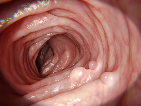
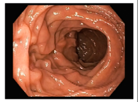
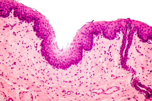
Free Images: "bestof:Tubulovillous Polyp of the Colon 1.jpg Tubulovillous Polyp of the Colon This polyp was removed by segmental sigmoid colon resection from an 18-year-old man who"
figures-statues-colon-sculpture-706871.jpg
Upper gastrointestinal tract.svg
Digestive system diagram numbered.svg
NonFreeImageRemoved.svg
Digestive system diagram en.svg
Tubulovillous Polyp of the Colon 1.jpg
Villous adenoma of the sigmoid colon, gross pathology.jpg
Tubulovillous Polyp of the Colon 2.jpg
Adenomatous Polyp of the Colon 1.jpg
Adenomatous Polyp of the Colon 2.jpg
Abdominoperineal_resection.jpg
Diverticula, sigmoid colon.jpg
Villous adenoma of the sigmoid colon 1.jpg
Villous adenoma of the sigmoid colon 2.jpg
Villous adenoma of the sigmoid colon 3.jpg
Gray1076.png
Colon_Hospital_building.jpg
LLQ2unlabled.PNG
LLQnumbered.PNG
LLQlabled.PNG
Sobo_1906_399.png
Haeckel_Tetracoralla.jpg
Illu_lymph_chain09.jpg
Haeckel_Siphonophorae_59.jpg
Johann Georg v. Eckhart.jpeg.jpg
Daniel Chodowiecki.jpg
Caspar Aquila.jpeg.jpg
Christian Garve.jpeg.jpg
August Boeckh.jpg
Ernst Friedrich Germar.jpg
Paulus Gerhardt.jpg
Palmyrenian relief Louvre AO1556.jpg
Paul Eber.jpeg.jpg
Conrad Ekhof.jpeg.jpg
Johann Gottfried Eichhorn.jpeg.jpg
Joachim Heinrich Campe.jpeg.jpg
Benedikt Carpzov junior.jpg
Johann Franz Encke.jpeg.jpg
Sigmund Feyerabend (EESachse).jpg
Matthias Claudius.jpeg.jpg
Hermann Conring.jpeg.jpg
Nicolaus Copernicus Profil.jpg
Lucas Cranach senior.jpg
Friedrich Christoph Dahlmann.jpeg.jpg
Albrecht Dürer - HBürkner.jpg
Johann Veit Döll.jpg
Johann Gottlieb Fichte.jpeg.jpg
Paul Fleming.jpeg.jpg
Leopold I. von Anhalt-Dessau.jpeg.jpg
Johann Wilhelm von Archenholz.jpeg.jpg
Johann Arndt.jpeg.jpg
Ernst Moritz Arndt.jpeg.jpg
Johannes Aventinus.jpg
Johann Sebastian Bach.jpeg.jpg
Ludwig van Beethoven.jpeg.jpg
Friedrich Wilhelm Bessel - EESachse.jpg
Jakob Fugger (EESachse).jpg
Georg von Frundsberg.jpeg.jpg
Friedrich II. von Preußen.jpg
Gebhard Leberecht von Blücher.jpeg.jpg
Johann Friedrich Blumenbach.jpeg.jpg
Johann Elert Bode.jpeg.jpg
Jacob Böhme.jpg
Carl Friedrich Gauß.jpg
Christian Fürchtegott Gellert.jpeg.jpg
Salomon Gessner.jpeg.jpg
Johann Geiler von Kaysersberg.jpeg.jpg
Sebastian Brant.jpeg.jpg
Johann Gottlob Immanuel Breitkopf.jpg
Gregor von Brück.jpg
Gottfried August Bürger.jpeg.jpg
Martin Buzer (Bucer).jpg
Joachim Camerarius senior.jpg
Khoi_Women_and_Dutch_Colonist_WDL11267.png
Carl von Österreich-Teschen.jpg
Ferdinand I. von Österreich (HRR).jpg
Conrad Celtis (Protucius).jpg
Peter von Cornelius (1862).jpg
Ludwig Wilhelm von Baden-Baden.jpg
Johann Matthäus Bechstein.jpeg.jpg
August Hermann Francke.jpeg.jpg
Friedrich III. von Sachsen.jpg
Franz Joseph Gall.jpeg.jpg
Leopold von Braunschweig-Wolfenbüttel.jpg
Ferdinand von Braunschweig-Wolfenbüttel.jpg
Christian von Bunsen.jpg
Low_Country.jpg
Friedrich Wilhelm von Brandenburg.jpg
Curious_creatures_in_zoology_(15568638285).jpg
Franz II. von Österreich (HRR).jpg
Friedrich Wilhelm Bülow von Dennewitz.jpg
Konrad.von.Boyneburg.01.jpg
Low_Country_house_on_stilts.jpg
USS_America_visits_San_Francisco_141006-N-FR671-203.jpg
USS_America_homecoming_140915-N-FR671-240.jpg
USS_America_homecoming_140915-N-FR671-266.jpg
USS_America_commissioning_141011-N-FR671-532.jpg
USS_America_arrives_in_Valparaiso_140824-N-FR671-040.jpg
USS_America_arrives_in_Valparaiso_140824-N-FR671-049.jpg
Terms of Use   Search of the Day