Click Here for More Images from iStock- 15% off with coupon 15FREEIMAGES 
GABA B receptors  are G protein-coupled receptors, also called metabotropic receptors.  Binding of  an agonist (baclofen, red) leads to a G-protein coupled C-AMP signaling pathway. Source: PDB entries 7eb2, 6r3q,.
The three-dimensional structure of proteins is represented in a variety of conformations. This image is based on the ribbon structure of a protein. It is suitable for biology notebooks and book covers.\nThis image has not been created by AI.
3D cartoon and Gaussian surface models, chain id color scheme, PDB 7t3a, white background
A ball and stick model of Human Insulin. Isolated on black.
Nourish hair of shampoo or serum. Repair damaged hair concept, 3d rendering.
A conceptual image of an abstract network of multi-coloured organic strands arranged in a non regular pattern all connected together in a complex manner. The network is made from smaller multi-coloured squares and polyhedra, in vivid colours against a pastel toned background. Shows background and foreground elements to the structure.
Cells in the body examined by endoscope
Coronavirus, SARS‐Cov‐2 infects a humain cell, the virus enter by using a spike protein attache to a receptor on cells called ACE2
The glucagon like peptide-1 receptor (GLPr) is a G proteincoupled receptor (GPCR) that mediates the action of GLP-1, a peptide hormone. The activated receptor  has a strong effect  on the management of type 2 diabetes mellitus and obesity, including glucose homeostasis and regulation of gastric motility and food intake.
3D Rendered Illustration, visualization of Neurons firing neurotransmitters in the synaptic gap.
Cell membrane and biology, biological concept, 3d rendering. Computer digital drawing.
Artificial Intelligence Sustainable Responsible Technology
Cell membrane cholesterol is shown in pink. 3D cartoon and Gaussian surface models, PDB 7rpk, white background.
ATP Synthase Protein - 3d rendered image. Medical microbiology concept.  Structure and conformational states of the bovine mitochondrial ATP (Adenosine triphosphate) synthase. Chemical energy is synthesized in eukaryotic cells primarily. Hydrolase. Hologram view. HUD, FUI
Botulinum toxin (3d structure) which prevents the release of the neurotransmitter acetylcholine from axon endings at the neuromuscular junction. Infection with the bacterium causes botulism.
Evolution and Mystery of Life: Mollusks Living in Water
The calcitonin gene related peptide (yellow) binds to its receptor (blue) on neurons and smooth muscle cells of cerebral blood vessels, activating a signal cascade through G-proteins (dark blue) in this cells that  leads to a dilatation of the blood vessels (vasodilatation).
Phospholipid bilayer membrane structures that make up living cells and their contents. This is an image of membrane proteins in the membrane.\nThis image, which looks like a plastic model, evokes biology and biochemistry. It can be used as a cover for magazines, textbooks and presentation materials.\nThis image was not created by AI.
Nucleosome, molecular model. 3D illustration. The nucleosome consists of a DNA double helix wrapped around a core of histone proteins.
background
3D cartoon and Gaussian surface models, PDB 4cmh, white background
A ball and stick + ribbons model of beta-secretase - a protein that is important in forming healthy peripheral nerve cells, but is also involved in the onset of Alzheimer's disease.
3d rendered Gastric inhibitory polypeptide (GIP, glucose-dependent insulinotropic peptide) endocrine protein hormone, 3D illustration
Monkeypox Virus. 3D Render
HER2 (light blue)  locked in the inactive conformation by Trastuzumab (left) and Pertuzumab. Trastuzumab has drastically improved the prognosis of HER2-positive breast cancer patients.
Endosomes - 3d rendered image of Endosomes are membrane-bound organelles. They are parts of endocytic membrane transport pathway.  Endomembrane system in cells. Bilayer Lipid Membrane. Abstract medical illustration. Microscopic view. Microbiologic research concept.
Calcidiol molecule, also called calcifediol, major circulating metabolite of vitamin D3 produced in liver, 3D illustration
Covid-19 Coronavirus Variants Omicron Delta Gamma Beta Alpha Pandemic Virus Disease Mutation
3D cartoon and Gaussian surface models, chain id color scheme, PDB 3omv, white background
3d rendering of Easter eggs background
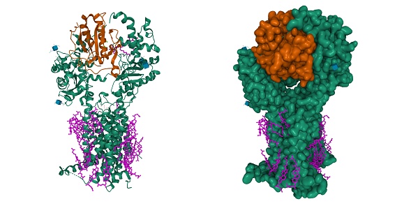
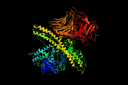
Free Images: "bestof:V receptor.png NMR structure of the intracellular loop i3 of the vasopressin V2 receptor GPCR PDB Jmol 2010-11-08 Jmol development team free screenshot GPL"
Conivaptan structure.svg
Mozavaptan structure.svg
Satavaptan structure.svg
A 276575 structure.svg
V receptor.png
Gprotein-coupled receptor kinase.png
Jmol2WUX.pdb.jpg
1ZS8.png
3JVF.pdb.jpg
4JVF.pdb.jpg
5JVF.pdb.jpg
PDB.png
2CZP.pdb.png
1IP0.pdb.png
1O8R.pdb.png
1BQS.pdb.png
1IVA.pdb.png
3B7X.pdb.jpg
3O5V.pdb.jpg
3LOY.pdb.png
3N7S.pdb.png
Galanin.png
Microcephalin.png
Nitrilase3IVZ.pdb.png
2KR6.pdb.png
3IVZ.pdb.png
Recoverin.png
Neurcalcin.png
Miraculin.png
Cd59.png
Cd59b.png
Kynureninase2HZP.jpg
3MSC.png
PDB1.png
Chimerinjmol.png
3P1M.pdb1.png
2W86 (Fibrillin).png
2VSZ.pdb1.png
3LL6.pdb1.png
Hexokinase.pdb1.jpg
Hexokinase.pdb2.jpg
Riboflavkinase.png
Factor D.jpg
2BC4.pdb1.png
1NEB.pdb.png
2KLL.pdb.png
1GPS.jpg
1H6K.pdb.jpg
Hexokinase 3O08.png
Fascin.pdb.png
Sramblase.png
Neuropilin.png
Cannabinoid receptor.png
1ZLL.png
EcdysonePhosphatePhosphatase.jpg
3L9S.pdb.jpg
1Z3U.pdb1.jpg
Pertussistoxin.png
2LNL.pdb.png
CGRP-receptor+RAMP1 3N7P.png
CFP-10.jpg
Kdesudo.png
Relaxin.png
Sorbitol dehydrogenase pymol image.png
VE template comparison.png
Mageia desktop.png
GNU Image Manipulation Program 005.png
GNU Health login popup.png
Sorbitol dehydrogenase pymol image 2.png
Sorbitol dehydrogenase pymol image-2.png
Os cust highlight.PNG
Rhodopsin 3D.jpeg
Quantaplus.png
Mouse 5HT3 receptor structure.png
Steroid receptor.png
3RZE-human histamine H1 receptor-pdb.jpg
Beta-2 adrenergic receptor.png
ANP+receptor 1YK0.png
BNP+receptor 1YK1.jpg
FSHA+B+receptor 1XWD.png
Human alpha parvalbumin.png
LDLR 1N7D.png
H1 Receptor with Doxepin.png
4GRV.png
Mozavaptan.png
SRX246 structure.png
3RZE.png
3V2Y.png
3VW7.png
Nicotinic Acetylcholine receptor.png
SeroNT.png
Met-Enkephalin 1PLW.png
Retrosharetransfer.png
NorepinephrineStructure.jpg
NAchR-2BG9 default.png
3UON.png
4DAJ.png
Protein Z.png
Protein structure 2K35.jpg
Jdownloader 0.8.png
Terms of Use   Search of the Day