Click Here for More Images from iStock- 15% off with coupon 15FREEIMAGES 
Stratified flat epithelium under microscope
Lymphocytic myocarditis. Myocarditis is an inflammation of the heart muscle. Lymphocytic refers to the type of cell seen in the affected tissue. A lymphocyte is a type of immune function cell.
Backgrounds of Bacteria and Mold under the microscope in Lab.
plant Pine Cone-Female L.S. under light mircoscope with white background
Microscopic preparation, tissue plant
Quantum Gravity 3d rendered image of a comprehensive exploration of Quantum Gravity theories. String theory, loop quantum gravity (LOG), causal dynamical triangulation, twistor theory, gravitational waves, spacetime, gravity waves, quantum mechanics, quantum entanglement concepts.
Epidermis. Electron microscope micrograph showing a keratinocyte of spinous layer. The epithelial cell has a polygonal shape, central nucleus with nucleolus, cytoplasm full of keratin filament bundles, and numerous dark desmosomes crossing the intercellular spaces.
Particles, medical concept
Leiden leaf c.s. under microscope
Photomicrograph of two rotifers. Rapidly rotating cilia at top of heads, internal organs visible. Live specimen. Wet mount, 10X objective, transmitted brightfield illumination.
Escherichia coli bacterium, E.coli, gram-negative rod-shaped bacteria, normal flora in GI tract and causative agent of diarrhea, close view.
Myxoid liposarcoma.  Myxoid/round cell liposarcoma, or MRCLS, is one of several types of liposarcoma. Liposarcoma is a rare cancer that grows in the cells that store fat in the body. MRCLS usually grows in the arms and legs. These tumors grow slowly, and they can spread to other parts of the body
Cross sections of the plant root under the microscope view for education
Slice of natural crystal for patterns and textures use with high resolution.
Host cells with spores (mold) are inside wood under the microscope for education.
Overhead aerial drone view of oyster farm racks at low tide, flying at a low altitude to be able to see the detail of the oyster racks and seaweed. Located in a tidal estuary in Sutherland, Scotland. the low tides enable workers to access the racks for turning the oysters as they grow. Oysters have been farmed for many hundreds of years using this method. Seaweed has grown around the metal lines of racks.
Transmission electron microscope (TEM) micrograph showing dark melanin granules in a melanocyte (top center) and in several keratinocytes, which, unlike melanocytes, have bundles of keratin filaments
Colourful abstract background
3D cartoon and Gaussian surface models, chain entity color scheme, PDB 4oo8, white background
Neuroendocrine carcinoma well differentiated (carcinoid) rectum with immunostain synaptophysin
Photomicrograph of Histological analysis of Intramedullary SOL showing Astrocytoma, WHO grade-4
Photomicrograph of flatworm,
Hyaline cartilage, Elastic cartilage and Bone Human under the microscope in Lab.
Bacterial multidrug efflux pumps (E-CmeB) expel antibiotics  from inside the bacterium (bottom): ciprofloxacin (yellow), chloramphenicol (violet),  ampicillin (red).
Yellow abstract background
Zona fasciculata of the adrenal gland. 0.5 µm thick semithin section of plastic-embedded material stained with toluidine blue. One of the most striking characteristics of the steroid-secreting cells of the adrenal cortex is the presence of abundant lipid droplets in their cytoplasm.
Focal nodular hyperplasia (FNH) is a benign liver lesion that is composed of a proliferation of hyperplastic hepatocytes surrounding a central stellate scar. Typically, FNH is a solitary lesion that is more commonly seen in women.
Microscope photo of a sambucus stem with parenchyma cells.
\
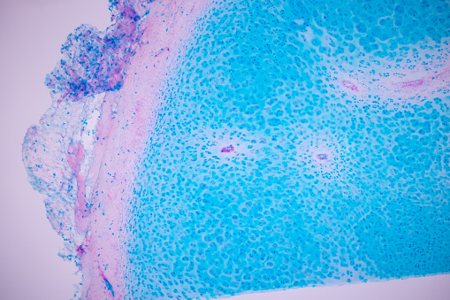
Free Images: "bestof:Vaginal wet mount with clue cells.jpg Vaginal wet mount with a NaCl preparation showing clue cells among normal epithelial cells Taken with light microscopy at"
3._Red_blood_cells_showing_signs_of_yeast.jpg
1._Healthy_red_blood_cells.jpg
Harmful_Water_Organisms.jpg
Villarceau circles.gif
red-light-cells-allianz-arena-1636281.jpg
light-solar-cells-lamps-hell-9181.jpg
light-solar-cells-lamps-hell-9182.jpg
2._Unhealthy_red_blood_cells.jpg
Mountain-Artist-Ridge-Trail-Park-Mount-Baker-Washington.jpg
source-jet-drops-of-water-1444348.jpg
Nude mouse (1).jpg
Osmia_texana,_female,_back,_Carroll_County,_MD_2013-10-30-14.32.30_ZS_PMax.jpg
Osmia_texana,_female,_side,_Carroll_County,_MD_2013-10-30-14.41.44_ZS_PMax.jpg
bee_19992,_f,_angle,_kenya_2014-08-07-14.44.23_ZS_PMax.jpg
bee_19992,_f,_face,_kenya_2014-08-07-14.58.57_ZS_PMax.jpg
Design_Analysis_of_the_Lockheed_PV-1_Ventura.png
Design_Analysis_of_the_F6F-5_Hellcat.png
portugues-sexo_alemão-sex_ingles-sex_africano-seks_russo-секс_albanes-sex_chines-性別_bulgaro-секс_croata-spol_gales-rhyw_coreano-섹스_frances-sexe_japones-セックス_filandes-sukupuolesta_arabe-الجنس_prontos_ai_tens_em_15_linguas_a_p.jpg
Design_Analysis_of_the_B-24J_Liberator.png
PHYSIC_STREET,_CANTON.jpg
Cellular tight junction-en.svg
Cellular tight junction keys.svg
Cellular tight junction-sv.svg
Cellular tight junction-fr.svg
Cellular tight junction-it.svg
PMID12529460 01.svg
PMID12529460 02.svg
Design_Analysis_of_the_P-38_Lightning.png
Lincoln_and_generals_at_Antietam.jpg
microscope-cells-laboratory-science-29520.svg
Mitosis cells sequence.svg
nerves-cells-dendrites-sepia-346928.jpg
nerves-cells-dendrites-sepia-791393.jpg
honeycomb-pattern-hexagon-design-34984.svg
background-structure-pattern-nerves-826334.jpg
background-tile-abstract-texture-1336607.jpg
Reference ranges for blood tests - blood cells.svg
SimpleCircuit.SVG
Mitotic Cytokinesis.svg
Zeppelin-LZ-127 internal and gas cells.svg
microscope-microscopy-science-294185.svg
Zeppelin-LZ-127 internal and gas cells opaque.svg
Mitotic Prophase.svg
Mitotic Prometaphase.svg
Mitotic Metaphase.svg
Mitotic Anaphase.svg
Mitotic Telophase.svg
Peroxisome.svg
Peroxisome (zh-cn).svg
Zinc-carbon-english.svg
Zinc-carbon-numbered.svg
Animal mitochondrion diagram en (edit).svg
Animal mitochondrion diagram unlabelled.svg
LloydsMethod3.svg
LloydsMethod1.svg
LloydsMethod2.svg
LloydsMethod15.svg
brakedance-break-dance-dancer-1250353.jpg
brakedance-break-dance-dancer-1242834.jpg
Oncogene activation illustration.jpg
Riassorbimento bicarbonati e respirazione cellulare.svg
Body centered cubic 35 balls.svg
View_of_Wonderland_showing_the_Royal_Hippodrome_and_Roller_Coaster,_Auckland_Exhibition,_Auckland_Domain.jpg
Outer hair cell and Deiter's cell.svg
Incandescent light bulb.svg
Sixth_plate_ruby_ambrotype_of_two_children_-_in_mount.jpg
Study-flow (zh-cn).svg
Study-flow.svg
Christian_and_Thompson_Figure_5_SVG.svg
Psychoacoustical tuning curves.svg
Peroxisome de.svg
Kathleen_Beyer_Blackburn_(1892-1968),_sitting_in_chair.jpg
Metastasis sites for common cancers.svg
Zeppelin-LZ-127 internal.svg
ShockleyQueisserFullCurve (DE).svg
Zeppelin-LZ-127 internal opaque.svg
ShockleyQueisserBreakdown2 (DE).svg
Design_Analysis_of_the_Boeing_B-17G_Flying_Fortress.png
Vaginal wet mount with clue cells.jpg
Vaginal wet mount with a clue cell.jpg
Vaginal wet mount of candidal vulvovaginitis.jpg
Vaginal wet mount in candidal vulvovaginitis.jpg
Detail of vaginal wet mount in candidal vulvovaginitis.jpg
Candida wet mount.jpg
Urine pus cells.jpg
ElodeaLeaffaceupcells.jpg
Bacterial vaginosis workup.jpg
Wiki A lumbricoides decorticated egg.jpg
Pearl_Bodies04.jpg
Eugène_Atget,_Junkyard_-_Getty_Museum.jpg
Platelet apheresis.jpg
Stool iodine and saline wet mount.jpg
H4IIE cells with stained nuclei.jpg
A lumbricoides fertilized egg.jpg
Pleomorphic.jpg
Chlorella with yellow indicative of chlorophyll.jpg
Pancreas FNA; adenocarcinoma vs. normal ductal epithelium (200x).jpg
Terms of Use   Search of the Day