Click Here for More Images from iStock- 15% off with coupon 15FREEIMAGES 
3D Render of a Topographic Map of Portland, Oregon, USA.
Adipose tissue human, Smooth muscles human, Bone human and Striated (skeletal) muscle human under the microscope in Lab.
Metastatic neuroendocrine tumor. Site: Liver.  Neuroendocrine tumors are cancers that begin in specialized cells called neuroendocrine cells. Neuroendocrine cells have traits similar to those of nerve cells and hormone-producing cells. Neuroendocrine tumors are rare and can occur anywhere in the body.
Product made after processing pictures taken from a drone. It shows forest area with a lot of trees and bushes. 3D rendering process allows to see height of a terrain while it looks like NIR photo.
Utah topographic map
Landscape topographic map
Abstract watercolor and acrylic flow blot smear painting. Blue landscape. Color canvas monotype texture horizontal background.
Bubbling water in a hot tub whirlpool
Snowcapped mountains in Colorado. Picture taken from high above in a plane during the winter.
3D Render of a Topographic Bathymetry Map of the Gulf of Mexico. \nAll source data is in the public domain.\nColor texture: Made with Natural Earth.\nhttp://www.naturalearthdata.com/downloads/10m-raster-data/10m-cross-blend-hypso/\nWater texture: SRTM Water Body SWDB: https://dds.cr.usgs.gov/srtm/version2_1/SWBD/\nRelief texture: ETOPO 2022 data courtesy of USGS. URL of source image:\nhttps://www.ncei.noaa.gov/products/etopo-global-relief-model
folded maps isolated on white
Cardiac amyloidosis is a condition where faulty proteins build up in your heart. You can inherit this condition, or it can develop on its own (usually later in life). As faulty proteins accumulate, your heart struggles to pump, ultimately leading to heart failure and death.
Product made after proccesing pictures taken from a drone. It shows excavation site with steep rock walls
Abstract landscape with horizontal lines and glowing lights
Aerial view of river meander in the lush green vegetation of the delta.\nBeautiful landscape - wild river in USA.\nNational nature reserve in summer.
Commercial photography of Crystal material background.
False colour TEM micrograph. Olfactory nerve showing the ensheathing glia (nucleus, blue, cytoplasm green, basal lamina, pink) surrounding many non-myelinated axons (brown) of olfactory neurons.\
Reed Islets in clear lake
Hyaline cartilage, Elastic cartilage and Bone Human under the microscope in Lab.
3D Render of a Topographic Map of Hamburg, Germany.
Salivary mucoid cyst. microscopic show parotid tissue, features of cyst wall composed of myxoid material and histiocytes. Histology. Parotid cancer.
Aerial View of Wetland, Izmir
Stowe, Vermont, Lamoille County, VT
Microscopic photo of a professionally prepared slide demonstrating gastric adenocarcinoma
Coloured transmission electron micrograph (TEM) of a small arteriole showing, from lumen: endothelium (magenta), discontinuous internal elastic lamina (blue), cross-sectioned smooth muscle fibers (cytoplasm, red, nucleus, light yellow) and connective tissue of adventitia (green).
There are so many burls and knot make the pattern like clouds with a beautiful nature and abstractive paint. High angle view of a flat textured wooden board backgrounds. A close-up studio shooting shows details and lots of wood grain on the wood table. The piece of wood at the surface of the table also appears rich wooden material on it. The wood is Saint Patrick green color with black lines and pattern on the surface. Flat lay style. It’s high-resolution textured quality.
Hipsometric model created by 3d rendering of photos taken from a drone. It shows forest area with a lot of trees.
Aerial view of dramatic landscape of Sankoh Prairie(桑科草原)
Blue toned Satellite Image of Rome, Italy. Digital Composite.  Contains modified Copernicus Sentinel data (September 03, 2016) courtesy of ESA. URL of source image: https://scihub.copernicus.eu/dhus/#/home. The source data is in the public domain.
Tissue of Small intestine (Duodenum), Large intestine Human and Stomach Human under the microscope in Lab.
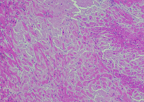
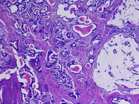
Free Images: "bestof:Vestibular schwannoma topography.jpg en Topography of vestibular schwannoma upper picture - normal condition Picture was scanned from book 1957 year Due to"
Terms of Use   Search of the Day