Click Here for More Images from iStock- 15% off with coupon 15FREEIMAGES 
background
Neon molecular atomic structure background. Round purple connected particles with 3d render abstract streaks in space. Volumetric futuristic biochemical and scientific formula
Cell membrane structure background, 3d rendering. Digital drawing.
A model of a molecule of estriol, one of several estrogen steroid hormones produced by the sex glands in mammals.  Isolated on white.
3D cartoon and Gaussian surface models, chain id color scheme, PDB 7t3a, white background
Molecular model of amylose, one of the two sugar components in starch
Cell Membrane Phospholipid Structure,Phospholipid bilayer of the cell membrane,Lipid bilayer
Chain of amino acid or bio molecules called protein - 3d illustration
Bacterial multidrug efflux pumps (E-CmeB) expel antibiotics  from inside the bacterium (bottom): ciprofloxacin (yellow), chloramphenicol (violet),  ampicillin (red).
Metastatic melanoma. Site: Neck. Metastatic melanoma is a disease that occurs when the cancerous cells from the original tumor (primary tumor) get loose, spread by traveling through the lymph or blood circulation, and start a new tumor (metastatic tumor) somewhere else.
DNA close-up, DNA Molecule Model, DNA Strands, DNA animation
Liver Fluke Section under light microscope with white background
3D cartoon and Gaussian surface models, chain id color scheme, PDB 3omv, white background
Molecular model of lactose, a disaccharide sugar in milk.
Proteasomes are molecular machines for breaking down proteins called proteolysis, only target proteins that have been marked for destruction called ubiquitin to the target protein, 3d rendering
Shiny artificial molecular structure in many colors. Horizontal composition, isolated on black background.
Thyroid cancer: Microscopic image of Metastatic papillary carcinoma of thyroid. Reactive change. Lymph node carcinoma.
Focal nodular hyperplasia (FNH) is a benign liver lesion that is composed of a proliferation of hyperplastic hepatocytes surrounding a central stellate scar. Typically, FNH is a solitary lesion that is more commonly seen in women.
PAPS Smear study of a young women under microscopy showing Atrophic changes in uterus which is unusual at young age.
A microscope slide showing a stained section of a human bowel, or small intestine.  It was taken through microscope lens that has a central focus through a thin layer of glass.
3D cartoon and Gaussian surface models, PDB 1uea, white background
\
3d rendering of compromised liposome releasing its contents. Magnified image of a liposome, showing leakage of its encapsulated material.
Bubble galaxy, an abstract image of high magnification soap bubbles with food dye to add interesting color. Studio shot with back illumination through translucent pan of soap bubbles.  Arty image that looks like colorful deep space.  Useful for abstract backgrounds and textures.
Kidney cancer: Clear cell carcinoma. fuhrman nuclear grade-II, Renal growth shows a malignant tumor, renal cell carcinoma in between renal tubules.
anaplastic thyroid carcinoma with heterologous differentiation (leiomyosarcoma)
ribonicleic acid chain from which the deoxyribonucleic acid or DNA is composed - 3d illustration
Molecular model of amylose
3D cartoon and Gaussian surface models, PDB 6vg8, white background
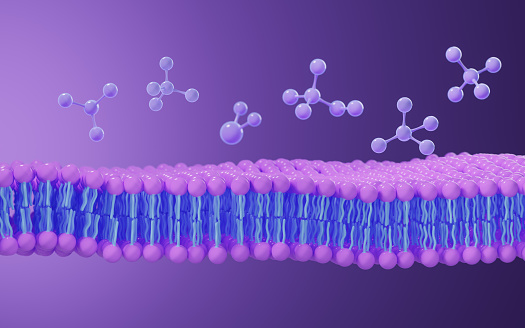
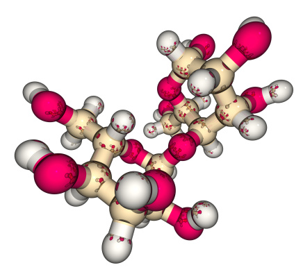
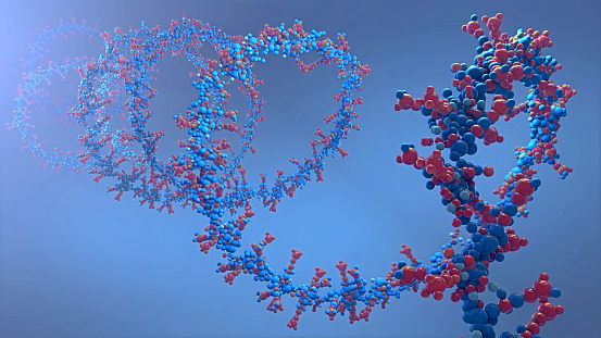
Free Images: "bestof:Lipid Molecules (5941036454).jpg The purple tails of the lipid molecules that form the cell membrane are far less orderly in the absence of cholesterol top"
Lipid vesicle vs soap bubble.svg
Cell membrane detailed diagram blank.svg
Cell membrane detailed diagram en.svg
Pore schematic.svg
Cell membrane detailed diagram id.svg
Lipid raft organisation scheme.svg
Black lipid membrane.svg
portugues-sexo_alemão-sex_ingles-sex_africano-seks_russo-ÑекÑ_albanes-sex_chines-性別_bulgaro-ÑекÑ_croata-spol_gales-rhyw_coreano-섹스_frances-sexe_japones-セックス_filandes-sukupuolesta_arabe-الجنس_prontos_ai_tens_em_15_linguas_a_p.jpg
Supported bilayer.svg
Bilayer hydration profile.svg
Scheme facilitated diffusion in cell membrane-en.svg
Lipid vesicles.svg
Scheme facilitated diffusion in cell membrane-zh.svg
Scheme facilitated diffusion in cell membrane-ca.svg
Scheme facilitated diffusion in cell membrane-eo.svg
Scheme facilitated diffusion in cell membrane-fr.svg
Scheme facilitated diffusion in cell membrane-zh (zh-tw).svg
Lipid bilayer fusion.svg
Endocytosis types-fr.svg
Endocytosis types.svg
Cell membrane detailed diagram fr.svg
Lipid unsaturation effect.svg
Lipid unsaturation effect pt.svg
Cell membrane detailed diagram ca.svg
Scheme simple diffusion in cell membrane-en.svg
Scheme simple diffusion in cell membrane-de 02.svg
Lipid unsaturation effect es.svg
Scheme simple diffusion in cell membrane-es.svg
Scheme simple diffusion in cell membrane-de.svg
Scheme simple diffusion in cell membrane-zh (zh-cn).svg
Scheme simple diffusion in cell membrane-pl.svg
Scheme simple diffusion in cell membrane-fr.svg
Scheme simple diffusion in cell membrane-ca.svg
Scheme simple diffusion in cell membrane-zh (zh-tw).svg
Animal mitochondrion diagram en (edit).svg
Endocytoza typy.svg
Complement pathway.svg
Pinocytosis.svg
Lipid Molecules (5941036454).jpg
Anesthesia (7003164721).jpg
Lipid bilayer expansion hypothesis of anesthetic effect.png
Cell membrane detailed diagram-el.png
Fluidní mozaikový model bunÄ›Äné membrány.tif
CellMembraneDrawing sk.jpg
LDLR pathway.png
Bicelleproteinmembrane.jpg
Bilayer AFM schematic.png
Space-Filling Model Sphingomyelin and Cholesterol.jpg
HLA-A1.png
CETP 2odb.png
Sedimented red blood cells.jpg
Lipid bilayer section.gif
Nanopore-based single molecule mass spectrometry (5884864158).jpg
Liposome.JPG
Membrane fusion via stalk formation.jpg
G protein1.png
NIST Membrane Model (5880464365).jpg
Crowded cytosol.png
DiffusionMicroMacro.gif
Endocytosis types (id).jpg
Scheme simple diffusion in cell membrane-he.png
Mixed lipid corral.jpg
Scheme simple diffusion in cell membrane-de.png
Putative mechaism of action of Human IRGM.jpg
PDB-7TM3.png
Interpretation of the cutoff effect in the frame of lipid hypothesis of general anaesthesia.jpg
Complement pathway.png
Junctional complex and pinocytotic vesicles - embryonic brain-TEM.jpg
A2-peptide.PNG
Unctional complex and pinocytotic vesicles - embryonic brain - TEM.jpg
F._W._Bolgiano,_seedsmen_have_won_confidence_for_ten_years_(16186336130).jpg
Bolgiano's_1902_catalogue_(15727587274).jpg
Design_Analysis_of_the_Vought_F4U-1D_Corsair.jpg
Terms of Use   Search of the Day