Click Here for More Images from iStock- 15% off with coupon 15FREEIMAGES 
Fruiting bodies (conidiophores) and hyphae of the fungus Aspergillus niger. A. niger is a widely distributed saprophyte which grows on household dust, soil, and decaying vegetable matter, including stale food. A. niger is one of the most common causes of fungal ear infections and may result in the serious lung disease aspergillosis in people with compromised immune function or other lung diseases. A. niger is used for waste management and biotransformation in addition to other industrial uses, such as production of citric acid and extracellular enzymes. Coloured scanning electron micrograph (SEM), magnified x415 when printed at 10cm wide.
Night landscape, star trails over the mountains.taken in Chadan Wetland, Zaduo County, Qinghai Province, China in August 2022
Ferrochromium (ferrochrome) piece on black background. Used for stainless steel production. Produced in South Africa, China, Kazakhstan, India, Russia, Finland. Copy space.
Spherical flower ,Monochrome
Abstract blue dot and line connection, Technology abstract background, Digital cyberspace background 3d Rendering
Blue fiber optics. XXXL!
Monosodium glutamate, MSG or sodium glutamate molecules. MSG is used in cooking as a flavor enhancer with an umami taste 3d rendering
Surface photography of nature pattern Crystal shot for background.
Abstract micrograph of ascorbic acid crystals in a bewildering array of shapes and colors.
Magnesium Lithium Aluminum Battery Element Mineral Metal Alloy
Leiden leaf c.s. under microscope
Quantum Gravity 3d rendered image of a comprehensive exploration of Quantum Gravity theories. String theory, loop quantum gravity (LOG), causal dynamical triangulation, twistor theory, gravitational waves, spacetime, gravity waves, quantum mechanics, quantum entanglement concepts.
This Tourmaline photographed with the macro in best studio quality and high resolution
Pegmatite rock close up with many crystals.
The Night Sky at Puertecillo Forests. Puertecillo is a small village close to Santiago de Chile where you can find nice beaches, forests and an amazing nature. During the night the view of the stars is awesome
A banded Agate stone sample with crystals texture.
Electricity fire-ball. Abstract photo of electric waves. Static electricity
Umbria, Italy: High voltage pylons
Microbacterium Tuberculosis Bacteria (MTB) in sputum smear AFB stained analysed by light microscope. Close up micrograph
Rural power lines cross a vast field under a deep blanket of snow covering Nova Scotia in February 2017.
telecommunication mast TV antennas wireless technology
A long , 30 minute , exposure of the sky at might with planes, going into Manchester Airport, Star trails, revolving around the pole star ,  and a few meteors from The Perseid meteor shower,
Plant tissue Structure, section (tissue) of stem plant tissue under a light microscope.
Abstract background of wires and glowing particles
Holographic iridescent 3d object, line gradient Holographic background
Dengue virus cell. 3D Render
Industrial quadrocopter performs work in the energy sector
Mineral Fusinus sp. Pliozän
Abstract image. Space, atoms, planets, molecules, electrons on a black background.
Starling Mumuration Black & White
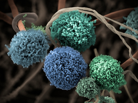
Free Images: "bestof:Magnetic fields; Transmission electron microscope (5884978267).jpg Transmission electron microscope TEM images show sections of a film of nickle-iron-copper-mol..."
Butterfly tongue.jpg
Leaf epidermis.jpg
Misc pollen.jpg
cells-electron-microscope-macro-570676.jpg
bacteria-electron-microscope-811861.jpg
bacteria-electron-microscope-808154.jpg
bacteria-electron-microscope-stained-808158.jpg
pollen-microscope-543490.jpg
hela-cells-cultured-544312.jpg
salmonella-bacteria-macro-549608.jpg
citrus-yellow-mite-macro-989500.jpg
cancer-cells-cells-scan-541954.jpg
pestis-bacteria-bubonic-plague-541948.jpg
blood-cells-cells-human-543484.jpg
bacteria-disease-escherichia-coli-62993.jpg
mite-aceria-anthocoptes-acari-67638.jpg
white-blood-cell-cell-blood-cell-543471.jpg
koli-bacteria-escherichia-coli-123081.jpg
yellow-mite-macro-lorryia-formosa-847873.jpg
yellow-mite-macro-lorryia-formosa-847862.jpg
Misc pollen colorized.jpg
Colletes_validus,_female,_face_2012-04-12-15.30.jpg
Colletes_validus,_male,_side_2012-04-12-12.46.jpg
3C58:_A_supernova_remnant_10,000_light_years_from_Earth..jpg
Harmful_Water_Organisms.jpg
Coax and Skin Depth.svg
Signale REM.svg
Wireless power - resonant inductive coupling.svg
Pear interaction SEM german.svg
Elaine_Zworykin.jpg
Hepatitis B virus v2.svg
Em fields field (physics).svg
Microscope optique simplifie principe.svg
Magnetic fields; Transmission electron microscope (5884978267).jpg
Magnetic fields; Transmission electron microscope (5884978021).jpg
Bronchiolar area cilia cross-sections 1.jpg
Spindle centriole - embryonic brain mouse - TEM.jpg
Mitochondria - TEM.jpg
Eosinophil TEM.jpg
Pancreatic cells - TEM.jpg
Two desmosomes - TEM.jpg
Chlamydomonas TEM 01.jpg
Chlamydomonas TEM 02.jpg
Chlamydomonas TEM 03.jpg
Chlamydomonas TEM 04.jpg
Chlamydomonas TEM 05.jpg
Chlamydomonas TEM 07.jpg
Chlamydomonas TEM 06.jpg
Mitochondria, mammalian lung - TEM (2).jpg
Mitochondria, mammalian lung - TEM.jpg
Clara cell lung - TEM.jpg
Pancreatic acinar cells - TEM.jpg
Human jejunum microvilli 1 - TEM.jpg
Human jejunum microvilli 2 - TEM.jpg
Human jejunum microvilli 3 - TEM.jpg
Chlamydomonas TEM 08.jpg
Chlamydomonas TEM 09.jpg
Chlamydomonas TEM 12.jpg
Chlamydomonas TEM 13.jpg
Chlamydomonas TEM 15.jpg
Chlamydomonas TEM 16.jpg
Chlamydomonas TEM 17.jpg
Immotile cilia syndrome.jpg
Fibers of Collagen Type I - TEM .jpg
Chlamydomanas reinhardtii Flagella 5 - TEM.jpg
Chlamydomanas reinhardtii Flagella 6 - TEM.jpg
Fibers of Collagen Type I - TEM.jpg
Chlamydomanas reinhardtii Flagella 9 - TEM.jpg
Lung epithelium 80294-2.6.jpg
Human skeletal muscle tissue 1 - TEM.jpg
Human skeletal muscle tissue 2 - TEM.jpg
Human skeletal muscle tissue 3 - TEM.jpg
Macrophage in the alveolus Lung - TEM.jpg
Human leukocyte, showing golgi - TEM.jpg
Chlamydomonas TEM 17 with scale.jpg
Alveolar sac region of the lung - TEM.jpg
Mammalian pancreatic acinar cell, nuclear pores -TEM.jpg
A red blood cell in a capillary, pancreatic tissue - TEM.jpg
Desmosome - epithelial cell from mammalian lung tissue - TEM.jpg
Junctional complex and pinocytotic vesicles - embryonic brain-TEM.jpg
Unctional complex and pinocytotic vesicles - embryonic brain - TEM.jpg
Retino ME con sezioni.jpg
Golgi in the cytoplasm of a macrophage in the alveolus (lung) - TEM.jpg
Mammalian pancreatic tissue, nuclear pores and rough endoplasmic reticulum.jpg
Cilia.jpg
Vibrio cholerae.jpg
Transmission Electron Microscope Kemira.jpg
Transmission electron microscope Kemira.jpg
Electron Microscope-fr.png
Microvilli.jpg
Block Copolymer (15774427462).jpg
TEM; cobalt iron alloy (6436372317).jpg
Root-nodule01.jpg
First_TEM_at_ERRC.gif
Chemical Analysis; Magnesium Oxide Dice (5887269391).jpg
Austenite ZADP.jpg
Lorentz microscopy2.PNG
Magnetic lens.PNG
Astigmatism.PNG
TEM-HRTEM.jpg
Terms of Use   Search of the Day